Cerebellum
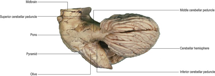
The cerebellum is the largest part of the hindbrain. It originates from the dorsal aspect of the brainstem and overlies the fourth ventricle. The cerebellum is connected to the brainstem by three stout pairs of fibre bundles, called the inferior, middle and superior cerebellar peduncles (Fig. 11.1); these join the cerebellum to the medulla, pons and midbrain, respectively. The functions of the cerebellum are entirely motor and it operates at an unconscious level. It controls the maintenance of equilibrium (balance), influences posture and muscle tone and coordinates movement.
External features of the cerebellum
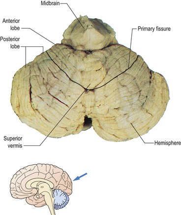
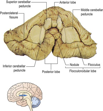
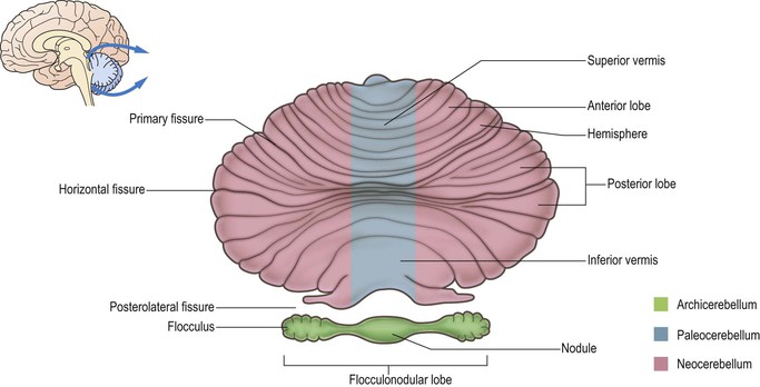
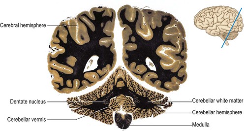
The cerebellum consists of two laterally located hemispheres, joined in the midline by the vermis (Figs 11.2–11.4). The superior surface of the cerebellum lies beneath the dural tentorium cerebelli and the superior vermis is raised, forming a midline ridge. Conversely, the inferior vermis lies in a deep groove between the hemispheres. The surface of the cerebellum is highly convoluted, the folds, or folia, being oriented approximately transversely. Between the folia lie fissures of varying depths. Some of these fissures are landmarks that are used to divide the cerebellum anatomically into three lobes (Figs 11.2–11.5). On the superior surface, the deep primary fissure separates the relatively small anterior lobe from the much larger posterior lobe. On the underside, the conspicuous posterolateral fissure demarcates the location of small regions of the hemisphere (the flocculus) and vermis (the nodule), which together form the flocculonodular lobe.

Internal structure of the cerebellum
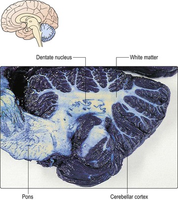
The cerebellum basically consists of an outer layer of grey matter, the cerebellar cortex, and an inner core of white matter. The white matter is made up largely of afferent and efferent fibres that run to and from the cortex, towards which it extends characteristic, irregular, branch-like projections. This formation is referred to poetically in older literature as the ‘arbor vitae’ or ‘tree of life’ (Figs 11.6, 11.7). Buried deep within the white matter are four bilaterally-paired cerebellar nuclei (Figs 11.6, 11.8), which constitute the output of the cerebellum to other levels of the neuraxis. They have important connections with the cerebellar cortex and with certain nuclei of the brainstem and thalamus.
< div class='tao-gold-member'>
Related posts:


Stay updated, free articles. Join our Telegram channel

Full access? Get Clinical Tree


