19 | Cervical Burst Fracture |
 | Case Presentation |
History and Physical Examination
A 24-year-old unrestrained driver was involved in a motor vehicle accident that required a prolonged extrication. He was urgently transported to a level I trauma center with appropriate spinal precautions. Physical examination revealed that he had no motor function distal to his trapezius bilaterally. He also had no sensation to pinprick distal to his shoulders. He had flaccid rectal tone and no sacral sensation. The patient was unable to maintain a mean arterial pressure above 70 mm Hg without inotropic support.
Radiological Findings
Imaging studies of the patient (Fig. 19–1, Fig. 19–2, and Fig. 19–3) revealed a C5 burst fracture and a C6 compression fracture. Magnetic resonance imaging (MRI) revealed C5-6 and C6-7 posterior interspinous ligament disruption with posterior longitudinal ligament transection at the C5-6 level with spinal cord signal changes consistent with edema and hematoma.
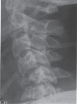
Figure 19–1 A lateral plain x-ray demonstrating a C5 and C6 vertebral body fracture with posterior element splaying resulting in a kyphotic deformity.
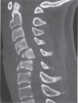
Figure 19–2 A sagittal computed tomographic reconstruction demonstrating a C5 and C6 vertebral body fracture with canal compromise at the level of C5.
Diagnosis
Based on his presentation and his imaging studies, a flexion compression stage V fracture of the C5 body with compression fracture of the C6 body was diagnosed. Retropulsion of the C5 body was present and the patient was noted to have a C4 American Spinal Injury Association-A (ASIA) neurological injury.
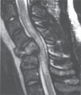
Figure 19–3 Sagittal T2-weighted magnetic resonance imaging demonstrating posterior longitudinal ligament disruption, C5-6 disk rupture and C4-5, C5-6 interspinous ligament injury.
 | Background |
According to the Allen-Ferguson classification, this particular patient has a flexion compression stage V injury to C5 due to retropulsion of vertebral fragments greater than 3 mm into the spinal canal and MRI evidence of posterior longitudinal ligament (PLL) and interspinous ligament disruption The C6 injury would be described as vertical compression stage II injury due to the anterior cortical failure resulting in breaking of the vertebral body with an associated fracture, although interestingly there is little loss of height at this level and absence of posterior ligamentous disruption.
The Allen-Ferguson system assumes one predominant force vector and one modifying vector based on neck position at the time of injury. This analysis results in six categories of injury: compression flexion, vertical compression, distraction flexion, compression extension, distraction extension, and lateral flexion injuries. Compression flexion injuries generally occur at the C4, C5, and C6 levels and involve failure of the anterior column in compression as well as the posterior elements in distraction. This pattern is subdivided into a spectrum of severity based on the cascade of injury from the anterior to the posterior column. Stage I and II injuries involve only the anterior column, whereas stage III involves the middle column. Stage IV and V injuries comprise failure in all three columns with advancing sagittal plane deformity. Vertical compression injuries occur most commonly at the C6 and C7 levels. The staging of these injuries ranges from the simple end plate cupping of stage I to stage III injuries, which involve bony failure with retropulsion. The key feature distinguishing flexion compression and vertical compression injuries is the lack of ligamentous failure in the latter. Distraction flexion, compression extension, distraction extension, and lateral flexion injuries are all subcategorized based on severity of inury.5
 | Authors’ Preferred Method of Surgical Management |
It is important to remember that all prospective spinal injury patients must undergo a spine series involving anteroposterior (AP) and lateral cervical, thoracic, and lumbar films as well as an open-mouth odontoid view. Any suspected spinal injury will require advanced spinal imaging. To better define the extent of bony injury, a computed tomographic (CT) scan with thin cuts and coronal and sagittal plane reconstructions is optimal. Additionally, magnetic resonance imaging (MRI) should be obtained when the patient is medically stable to evaluate spinal ligamentous, diskal, and spinal cord integrity. Finally, magnetic resonance angiography (MRA) may be indicated to rule out vertebral artery injury, especially if the foramen transversarium is violated, anticipated surgical instrumentation may risk injury to the vertebral vessels, or the cranial nerve exam suggests vertebral basilar insufficiency.1,2
In addition to the possibility of noncontiguous spinal injuries in up to 10% of cases of spinal trauma, vigilance for associated soft tissue injuries should be maintained. This, along with whole body “manscan” becomes even more imperative in the insensate patient. Once a spinal injury is diagnosed and other associated injuries are excluded or detected by the primary and secondary surveys, treatment of the spinal injury may commence. Although numerous studies have questioned its efficacy, intravenous high-dose methylprednisolone still remains the only known medicinal agent routinely administered less than 8 hours following a traumatic spinal cord injury. The current regimen employed at our institution is a 30 mg/kg methylprednisolone bolus followed by 5.4 mg/kg/h over 23 hours. The drip may be continued for 48 hours if initiated between 3 and 8 hours following injury or for 24 hours if initiated less than 3 hours after the injury.3,4 Appropriate medical management of high-dose steroid potential side effects is imperative. These include GI prophylaxis against gastrointestinal ulceration through use of sucralfate or proton pump inhibitors and blood glucose management with insulin. Preliminary traction to stabilize the fracture and improve alignment is performed with skeletal tong traction or a halo ring depending on the fracture type.
A halo ring traction may initially be applied as a reduction tool or as a stabilizing measure prior to operative management. Some centers may even consider halo vest immobilization following traction reduction as a definitive means of treatment for this injury type; however, the literature does not favor this approach in this particular case example.6–8 Surgery is heavily favored in this fracture subtype to improve the odds of long-term spinal stability and for improved spinal alignment. Spinal decompression for the sake of relieving pressure on the spinal cord in an ASIA-A patient may serve two functions: (1) to maximize the chance of expected single- or two-level cervical nerve root level recovery generally seen in these patients and (2) to improve cerebrospinal fluid (CSF) flow mechanics and possibly limit the late sequel of cystic degeneration of the spinal cord and subsequent further neurological deterioration.
In this case, the spine is circumferentially unstable and presents in a kyphotic posture in the sagittal plane; thus our treatment approach would be to improve sagittal alignment initially with closed skeletal traction up to ~40 to 50 lb followed by either temporary halo or collar immobilization prior to surgical stabilization. An anterior surgical approach would first be performed through a transverse incision. A C5 corpectomy would be performed using either autologous iliac crest bone or a fibula strut graft filled with autologous bone graft from the corpectomy bone as a bone graft source. Care to avoid involving C6 in the decompression if possible would greatly limit the extent of surgery and facilitate surgical reconstruction. Additionally, the preoperative MRI demonstrated increased signal intensity in the interspinous ligaments between C6 and C7. It was unclear if this would at a later time manifest as junctional instability if the fusion extent was limited to C6. Following the C6 corpectomy, a statically locked anterior cervical plate would be applied from C4 to C6. The patient is then carefully turned prone using a Mayfield tong head holder (Integra, Plainsboro, NJ). Using the standard technique of lateral mass screws, C4-7 posterior spinal fusion was performed, augmented by morcellized iliac crest bone graft or bone left over from the anterior corpectomy.
Our institution has gradually shifted away from autologous bone graft in nonsmoking trauma patients due to the associated bone graft harvest morbidities. In advanced-stage compression flexion injuries we prefer to perform a circumferential fusion to maximize fusion success. This is performed posteriorly with segmental lateral mass and pedicle screw fixation.9,10 Due to the presence of structural damage to the C6 body, it was questionable whether this vertebral body would be stable enough to accept an anterior screw. Fortunately this was the case and there was no need to perform an additional C6 corpectomy.
Pearls
Surgical induction should begin with fiberoptic intubation if the patient is not already intubated. This means of intubation is critical to avoid unnecessary cervical manipulation. In addition, the traumatized spinal cord is extremely sensitive to manipulation. It is not uncommon to have motor evoked potential or somatosensory evoked potential signal loss with even slight agitation of the spinal cord. Therefore, extreme caution should be taken during positioning and anesthesia, especially if the patient has an incomplete spinal cord injury. Mean arterial pressure would be invasively monitored and maintained at a mean pressure of ~90 mm Hg with IV pressor support if necessary throughout the surgical and perisurgical period to allow adequate spinal cord perfusion.


