Fig. 16.1
Left, PRESTIGE® ST cervical disc prosthesis; Right, PRESTIGE® LP prosthesis (Image provided by Medtronic, Inc)
The BRYAN cervical disc (Medtronic, Inc.) was designed by the American neurosurgeon Vincent Bryan from Seattle in 1990s. The concept and design of the BRYAN disc is completely different from the Bristol/PRODISC series. This device consists of two titanium alloy endplates articulating with a polyurethane core. The two titanium endplates are fixed to the bone by a porous titanium layer and stability is achieved through a tight fit of the prosthesis in the milled cavity (Fig. 16.2). The implant has been extensively tested in Europe and received US FDA approval in May 2009.
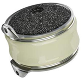
Fig. 16.2
BRYAN® Cervical Disc prosthesis (the BRYAN® Cervical Disc incorporates technology developed by Gary K. Michelson, MD. Image provided by Medtronic, Inc)
The third alternative to metal-on-metal implants is represented by the ProDisc-C device (Synthes, Inc.) which has recently obtained the approval for use in the United States. The ProDisc-C system was developed by Dr. Thierry Marnay in France and consists of two cobalt-chrome-molybdenum (CCM ) endplates with an UHMWPE articulating surface. It is a ball-and-socket constrained prosthesis and has a central keel for extra fixation in the vertebral body.
Other devices have recently joined the market of cervical TDR. Kineflex-C disc (Spinal Motion, Inc.) and CerviCore disc (Stryker Spine, Inc.) are metal-on-metal implants, whilst PCM (CerviTech, Inc.), DISCOVER (DePuy Spine, Inc.), and the MOBI-C (LDR, Inc.) are metal-on-UHMWPE implants.
Indications for Use and Contraindications
The rationale of considering TDR rather than a standard fusion procedure (i.e. ACDF) lies in the aim of preserving motion of the treated segment and preventing adjacent-segment degeneration. The typical candidate patient for TDR is the young active adult patient with single level symptomatic disc disease (i.e. radiculopathy) from C3 to T1 with intact posterior facet joints. General contraindications are marked reduction of the disc space with loss of motion at that level, zygapophyseal joint osteoarthritis, significant deformity in the sagittal and coronal plane, clear segmental instability, and infection. Other relative contraindications include rheumatoid arthritis, renal failure, osteoporosis, cancer, and preoperative corticosteroid use [20].
Evaluation of sagittal alignment, presence of zygapophyseal joint osteoarthritis and instability is of paramount importance and should be undertaken as routine preoperative assessment in every patient. Standard X-ray films (i.e. AP and lateral view) of the cervical spine and flexion-extension studies are usually sufficient in clarifying the extent of residual movement at the index level and the presence of osteoarthritic changes in the posterior joints.
The role of TDR in patients with axial neck pain has not been clarified yet and therefore disc pathology with no neurological symptoms should not be considered an indication for TDR. European and US trails have enrolled patients with cervical radiculopathy due to disc herniation (soft or hard), foraminal osteophytes as well as cervical myelopathy. In our clinical experience the presence of a hard disc herniation should be considered a relative contraindication to TDR due to frequent need of a more extensive disruption of the endplate for a satisfactory clearance of the canal. In both European and North American trials, there has been a strong prevalence of patients enrolled with radiculopathy (77–93 %) rather than cervical stenosis/myelopathy . The role of TDR in cervical myelopathy has been recently investigated by different authors. Sekhon and co-workers [21] from Australia reported on 11 patients with single level myelopathy treated with TDR and average follow-up of 18 months. Although significant improvement was reported in clinical outcome measures, two complications were noted. One patient developed heterotopic ossification, and another patient developed progression of myelopathic compression due to postoperative oedema . Moreover, worsening of sagittal alignment of the cervical spine was noted in three patients. On the other hand, other authors have reported their positive experience of TDR in myelopathic patients. Fay et al. [22] reported on the results of a comparative study of TDR in 151 consecutive patients with cervical radiculopathy and cervical myelopathy. At the average follow-up of 36 months, no differences were identified in the two groups in terms of clinical and radiographic outcomes . However in our opinion cervical TDR should be avoided in patients with cervical myelopathy. Complete clearance of the spinal canal and wide decompression of the spinal cord are top priorities in cervical myelopathy surgery and the achievement of a solid and stable fusion if the best single guarantee for a long term success of the decompression.
A summary of the most common indications and contraindications for cervical TDR is shown in Table16.1. Although a thorough discussion on the indications of TDR is not possible due to the recent introduction into clinical practice of this technique, indications and contraindications listed in the table are widely accepted by most authors.
Table 16.1
List of commonly accepted indications and contraindications to cervical total disc replacement
Indications for cervical TDR | Relative indications for cervical TDR | Contraindications for cervical TDR |
|---|---|---|
Radiculopathy caused by soft disc herniation | Radiculopathy caused by hard disc herniation | Osteoarthritis of the zygapophyseal joints |
Myelopathy caused by disc herniation | Sagittal malalignment of the cervical spine | |
Radiculopathy caused by foraminal osteophytes | Segmental instability | |
Infection | ||
Previous posterior surgery | ||
Ossification of the Posterior Longitudinal Legament (OPLL) |
Clinical Studies
BRYAN Disc
The BRYAN disc has the longest clinical and radiological follow-up among cervical TDR devices . The first multicentre study on this device was published in 2002 by Goffin and co-worker as part of a European prospective multicentre trial [23]. The study enrolled 60 patients with cervical radiculopathy or focal myelopathy non responsive to at least 6-weeks of conservative treatment. Exclusion criteria were the presence of sole axial neck pain, malalignment of the cervical spine, previous neck surgery and cervical instability. Only single level implants were used for this study and clinical success rates at 6 months and 1 year were 86 and 90 %. Because of the lack of a control group, the authors assumed from the literature a target level of success rate of 85 % for ACDF surgery. The number of patient lost at follow-up was significant with only 30 patients available at the 1-year follow-up. No complications directly related to the implant were detected. However, three patients underwent revision operation for prevertebral hematoma drainage, posterior foraminotomy for residual compression, and posterior laminectomy for residual myelopathy.
In a second study, Goffin and colleagues [24] expanded their original study with a second group of patients treated with two levels TDR. The study reported the results for 103 patients in the single-level group and 43 patients in the two-level group at 2 years follow-up. Success rates for the single-level group were 90, 86, and 90 % at 6 months, 1 and 2 years follow-up respectively. Patients in the two-level group had success rates of 82 % at 6 months, and 96 % at 1 year. No device failure or subsidence was reported in this second study and an average postoperative range of motion of 7.9° per level in flexion-extension was recorded. Movement was maintained in 87.8 % of the single-level patients and 85.7 % of two-level patients. Four complications were reported including one case of prevertebral hematoma, one case of epidural hematoma , one case of pharyngeal and oesophageal injury, and one case of residual nerve root compression.
Although enrolment criteria for the European study included patients with focal myelopathy, the actual number of patients with myelopathy enrolled in the study was minimal. In a separate study, Sekhon et al. [21] reported the results of BRYAN disc in treatment of 11 patients with cervical myelopathy with average follow-up from 1 to 17 months. No complications were reported and improvement of Nurick grade of 0.72 points and NDI scale of 51.4 points was noted. In contrast to these observations, Lafuente et al. reported on clinical results of 37 patients with cervical radiculopathy and 9 patients with cervical myelopathy . Analysis of the results showed that radiculopathy patients were doing better than myelopathy patients. Moreover, patients with myelopathy were also more likely to experience residual symptoms [25].
The first extensive report on North American experience with the BRYAN disc has been published by Sasso and co-workers in 2007 and 2008 [26, 27]. The authors conducted a prospective, three-center, randomized trial on 115 patients randomized in a 1:1 ratio to disc replacement and ACDF and plate surgery . Inclusion criteria were similar to the European studies and included patients with cervical radiculopathy and focal myelopathy due to single-level disc degeneration with symptoms non responsive to conservative treatment. Follow-up was 2 years for 99 patients. The authors reported a longer operative time for the arthroplasty group (1.7 h vs 1.1 h) but a significantly lower NDI for the disc replacement group at 12 and 24 months (11 vs 20, p = .005). Analysis of arm pain at 1 and 2 years also favoured the arthroplasty group with significantly lower VAS scores (14 vs 28, p = .014). The reported average range of motion per level in the disc replacement group was 7.9° in flexion-extension at 24 months, whilst it was 0.6° in the fusion group. No complications related to the implants were noted, as well as no heterotopic ossifications. Six patients underwent additional operations during the follow-up period, four patients in the control group and 2 patients in the BRYAN group. Four patients (2 in the control group and 2 in the BRYAN group) underwent a new ACDF surgery for adjacent segment degeneration .
The most recent and comprehensive study on BRYAN disc has been published by Heller and colleagues in 2009 [28]. This was part of the US IDE trial for FDA approval of the device and consisted of a prospective, randomized, controlled trial on 463 patients with minimum follow-up of 24 months. Inclusion criteria and outcomes measures were similar to the studies published by Sasso and co-workers [26, 27]. A total of 242 patients were enrolled in the BRYAN group and 221 patients in the control group (ACDF with plating). Fusion occurred in 94.1 % of the ACDF patients at the final follow-up. Although both groups showed improvement of the outcome measures , analysis of the data favoured the BRYAN group in several outcomes, including NDI, neck pain and return to work. Overall success rate was 82.6 % for the disc replacement group and 72.9 % for the ACDF group at 2 years. Complications occurred in 75 patients (31.0 %) of the disc replacement group and 61 (27.6 %) of the fusion group. Almost all complication were related to general medical conditions, secondary procedures were needed in only 6 patients for the BRYAN group (1 revision, 3 removals of the implant, and 2 re-operations) and in 8 patients for the fusion group (3 removals, 1 re-operation, and 4 supplemental fixations). Total revision rate for the BRYAN group was 2.9 and 3.2 % for the ACDF group. The average range of motion at 24 months for the arthroplasty group was 8.1°.
ProDisc-C
The ProDisc-C implant has received the US FDA approval for use in single-level disc arthroplasty due to the good results reported by the IDE study by Murray and colleagues [29]. An earlier study by Bertagnoli et al. [30] reported on the results of 27 patients treated with single-level ProDisc-C implantation at 1 year follow-up. Patients experienced sustained improvement of their symptoms at 1 year follow-up with decrease of NDI and VAS scores. No device complications were reported.
The actual FDA approval study was published in 2009 [29]. It was a prospective, multicenter, randomized controlled trail conducted on patients with single-level pathology. A 1:1 randomization scheme was adopted, 106 patients were randomized into the ACDF group and 103 patients in the arthroplasty group. VAS, NDI, and SF-36 scores were recorded at 3, 6, 12, 18, and 24 months after surgery. Clinical outcome measures significantly improved in both groups after surgery and results were maintained at final follow-up. Arthroplasty group maintained range of motion at the index level in 84.4 %. Overall, the ProDisc-C group showed results equivalent or slightly superior to the ACDF group although there was a statistically significant difference in the complication rates. In the fusion group , 8.5 % of the patients needed re-operation, revision, or supplemental fixation compared with 1.8 % of the ProDisc-C group (p = .033).
PRESTIGE Disc
The Cummins/Bristol device was the precursor of the PRESTIGE series of disc arthroplasty. The Cummins disc was developed to address the problem of disc degeneration in patients with previous fusions or with Klippel-Feil syndrome. The first study on this device enrolled 20 patients and showed, at 5 years, significant clinical improvement and preservation of the movement in 88.9 % of the patients. Unfortunately, a high rate of complications was reported, including screw loosening, mobilization of the implant, dysphagia and transient hemiparesis.
The PRESTIGE I and II discs were developed as an evolution of the original Cummins disc. Clinical results of the PRESTIGE I disc were published by Wigfield and coworkers in 2002. A total of 15 patients were enrolled in a prospective non randomized trial. Inclusion criteria encompassed patients with cervical radiculopathy or single level myelopathy secondary to cervical disc herniation or foraminal osteophytes. No significant complications were reported by the authors and all patients showed preservation of motion at the index level at 2 years after surgery. Mean flexion-extension ROM was 6.5° and mean antero-posterior translation was 2 mm. Clinical improvement was documented by ODI, NDI, and SF-36 but no valuable statistical analysis was undertaken because of the small number of patients. The PRESTIGE II implant was studied by Porchet and Metcalf on 55 patients. Standard clinical and radiographic evaluation was undertaken by the authors and the results showed a substantial overlap between the artificial disc and the ACDF surgery group.
The best available data on clinical safety and efficacy of the PRESTIGE ST disc has been published in 2007 by Mummaneni and colleagues. Data from this report have also served as the basis for the current FDA approval of this device in the United States. The study consisted in a prospective 1:1 randomized trial with patients undergoing either single level disc arthroplasty or single level ACDF. A total of 541 patients were enrolled, 276 patients in the PRESTIGE ST group and 265 patients in the ACDF group. The study showed a two-point greater improvement of NDI in the investigational group at 12 and 24 months. Improvement in SF-36 questionnaire scores was higher in the arthroplasty group at 12 and 24 months, as well as the VAS score. The rate of revision surgery was lower for the interventional group (5 revision surgeries) vs the fusion group (23 revision surgeries). No device failures or complications were reported, the average motion preservation at 2 years was 7°. The PRESTIGE LP disc arthroplasty has received FDA approval for use in patients in July 2014.
In a recent meta-analysis, McAfee and colleagues have summarized best available evidences about the use of cervical total disc replacement in clinical practice. The authors looked at the reported results of four prospective randomized controlled FDA IDE trials using BRYAN, PRESTIGE, ProDisc-C, and PCM implants. Data from 1226 patients at 24 months were available for the analysis. Results showed an overall success rate of 70.8 % in the ACDF patients and 77.6 % in the arthroplasty group (p = 0.007), thus favouring this last treatment. The analysis of all clinical subcomponents (i.e. neck disability index, neurological status, and survivorship) also favoured arthroplasty over ACDF surgery at 24 months. Survivorship ranged from 90.9 % in the PRESTIGE group to 98.1 % in the ProDisc-C group. Survivorship was achieved by 96.6 % of the cervical arthroplasty group on average and by 93.4 % of the ACDF patients. Some criticism has been raised regarding the poor results of the ACDF surgery (70.8 % overall success rate) in the reported FDA IDE trials. As pointed out by the authors of the study a common perception of a much higher success rate in fusion patients undermines confidence in the results of these trials. FDA criteria for definition of success are much more stringent than what has been traditionally reported in observational studies on ACDF surgery. This may account for the lower than expected results of the control fusion groups; taken together these data suggest that cervical disc arthroplasty is at least as clinically successful as fusion at 24 months [31].
Complications
Cervical disc replacement surgery shares with standard anterior cervical fusion surgery the same risks related to surgical approach. In a recent retrospective review by Fountas et al. of 1015 cases of primary one, two, and three level ACDF and plating, reported mortality was 0.1 %; 9.5 % of the patients suffered from postoperative dysphagia, 3.1 % had recurrent laryngeal nerve palsy, 2.4 % prevertebral hematoma, 0.5 % had dural perforation, 0.1 % hardware failure, and 0.1 % wound infection [32]. Access related complications for cervical arthroplasty are in the same range. In the two European studies on BRYAN disc, 0.97 % of patients (1 out of 103) required evacuation of prevertebral hematoma, and 2.91 % (3 out of 103) required additional surgery to decompress the neural canal. Dural tear was noted in 2.33 % of patients, and one patient required oesophageal tear repair [23, 24]. Analysis of complications in one FDA IDE trail showed more general medical complications in patients who underwent total disc replacement than the fusion group. Dysphonia/dysphagia was noted in 10 % of patients in the arthroplasty group, and 2.8 % of patients developed wound infection [28].
Related posts:


Stay updated, free articles. Join our Telegram channel

Full access? Get Clinical Tree


