Overview
Disorders of the cervical spine can cause radiculopathy, myelopathy, or both. Compression of the neural elements occurs most commonly as a result of disk herniation and/or osteophyte formation but can also be caused by congenital deformities, facet joint hypertrophy, infection, and neoplasm. Conservative, nonoperative management is initially recommended for most patients with radiculopathty.
In patients refractory to nonoperative treatment, surgical intervention can often lead to significant long-term improvement in quality of life. Depending on the etiology, posterior cervical microforaminotomy (PCMF) or decompressive laminectomy can offer several important benefits over an anterior cervical approach and fusion: better preserved neck motion, no complications associated with instrumentation, no risk of pseudarthrosis, and decreased costs associated with shorter operative times and lack of implants. Posteriorly approached tandem foraminotomies have similar outcomes while maintaining superior neck motion when compared with anterior decompressions and fusions. Multilevel disease, even with ventral spinal cord compression, may be adequately decompressed with cervical laminectomy. Reports have suggested that adjacent-segment disease and C5 nerve root palsy are also mitigated by the posterior approach.
In patients with sagittal or coronal deformity or any signs concerning for instability, a concomitant fusion may be appropriate and unavoidable. Furthermore, if the majority of the disk or facet joints are removed, the posterior decompression itself can lead to instability.
Diagnosis
As is normally the case, patient anamnesis is critical for making the correct diagnosis and for surgical planning. Typically, patients come to medical attention with complaints of neck and radiating arm pain. This is often associated with numbness or tingling in the arms and fingers. In addition, they may indicate difficulties with specific tasks, such as opening a jar, playing a guitar, or turning a car key. It is similarly important to inquire about clumsiness and gait and balance issues, as these can be early signs of myelopathy.
Radiographic images—such as magnetic resonance imaging (MRI) scan or computed tomographic (CT) myelogram, the latter being useful in patients with previously instrumented fusion—should supplement, not replace, the patient history and exam ( Fig. 20-1 ). Plain radiographic images including anteroposterior (AP), lateral, and flexion and extension views, should always be obtained to evaluate spinal curvature, alignment, and motion. Oblique views may be helpful in assessing foraminal stenosis. When the source of the pain is difficult to localize, selective nerve root blocks or electromyography with nerve conduction velocity testing may be considered.

The goal of surgery may dictate the approach taken and the number of cervical levels addressed. Surgery may be limited to address symptoms or may extend more broadly to address subtle signs and radiographic abnormalities. The patient must be included in the decision-making process and must be informed of all advantages and disadvantages associated with any given intervention.
Indications and Contraindications
Posterior Cervical Microforaminotomy
Indication
- ▪
Cervical radiculopathy at one or more levels caused by disk herniation and/or osteophyte formation
Relative Contraindications
- ▪
Cervical myelopathy
- ▪
Midline disk herniation
- ▪
Cervical instability at the pathologic level
- ▪
Preoperative cervical kyphosis
- ▪
Vertebral body pathology
- ▪
Disk herniation with bilateral radiculopathy at the same level
Laminectomy
Indications
- ▪
Multilevel cervical spondylotic myelopathy
- ▪
Cervical stenosis involving three or more levels
- ▪
Ossified posterior longitudinal ligament (OPLL) at multiple levels
- ▪
To access an intradural pathology (e.g., extramedullary and intramedullary neoplasm)
- ▪
Structural difficulties with an anterior approach (previous anterior surgery, short neck, barrel chest, obesity)
- ▪
Failure of previous anterior decompressive surgery
Contraindications
- ▪
Kyphotic deformity
- ▪
Instability of the pathologic level
- ▪
Younger patients (high risk of developing kyphotic deformity)
Operative Technique
Equipment
The Mayfield head clamp is recommended over a horseshoe headrest to avoid pressure on the central retinal artery. Excessive and extended pressure on the orbits and central retinal artery can lead to blindness.
Patient Positioning
PCMF or decompressive laminectomy is best approached with the patient in a prone position. The sitting position may reduce blood loss secondary to collapse of the epidural vessels but requires greater preoperative preparation and intraoperative vigilance for detection of air emboli. The embolic risk in prone position is relatively small and can be further reduced by proper positioning. The patient’s knees should be positioned higher than the heart, and bilateral compression stockings should be the standard of care. The authors prefer and recommend the prone position with the patient’s chin flexed approximately 45 degrees to reduce cervical venous pressure and to increase the interlaminar space. With the patient secured, the reverse Trendelenburg position is often used so that the cervical spine is roughly parallel to the floor ( Fig. 20-2 ).

Minimally Invasive Approach
With the minimally invasive approach to PCMF, a spinal needle is inserted approximately 1 to 2 cm off midline at the pertinent level. The target, the junction of the medial facet joint and two laminae, should then be confirmed with intraoperative fluoroscopy. A 12- to 14-mm stablike incision is made over the needle puncture site and is followed by removal of the spinal needle and insertion of a guidewire or a small dilator. The dilator is preferred over the guidewire to avoid penetration of the ligamentum flavum and creation of an inadvertent durotomy.
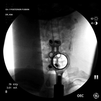
The dilator should be advanced and docked on the corresponding lateral mass. This can be confirmed by intraoperative fluoroscopy. Gentle soft-tissue dissection is carried out with subsequent dilators until the appropriate sized tubular retractor can be inserted. The location should again be confirmed by intraoperative fluoroscopy ( Fig. 20-3 ). The operative microscope is then brought over the surgical field to allow for direct visualization of the corresponding laminae and facet joint. The subsequent steps, whether done via a minimally invasive approach or by open PCMF, are identical and are described below.
Related posts:
 Craniovertebral Junction Instabilities and Surgical Fixation Techniques
 Surgical Approaches to the Craniovertebral Junction in Rheumatoid Arthritis
 Thoracic Microdiskectomy: Lateral and Posterolateral Approaches
 Lumbar Facet Screw Fixation Techniques
 Surgical Management of Scheuermann Kyphosis
 Primary Malignant and Benign Tumors of the Spine
Stay updated, free articles. Join our Telegram channel

Full access? Get Clinical Tree


