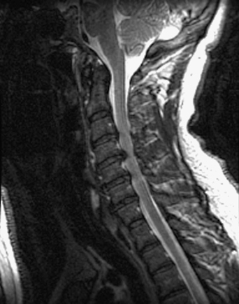
29 A 44-year-old man with chronic neck pain began experiencing hand numbness. On examination he was myelopathic. A T2-weighted sagittal magnetic resonance imaging (MRI) of the cervical spine revealed C3 through C6 ventral cord compression (Fig. 29-1). Cervical spondylomyelopathy A 360-degree cervical fusion, with a three-level corpectomy, titanium cage, and plating anteriorly, and a lateral mass fixation posteriorly (Figs. 29-2 and 29-3) were done. Several factors are considered in determining surgical approach when faced with cervical spondylomyelopathy. These include cervical contour, presence or absence of kyphosis, the nature and length of the compression, the patient’s overall condition and bone quality, and the surgeon’s experience with a given approach. This patient presented with significant myelopathy at a relatively young age. Because of his ventral cord compression, a multilevel corpectomy was needed to sufficiently decompress the cord. There are several long-arm corpectomy graft options. Autograft is a safe and successful option, but obtaining one to fit a three-level corpectomy can be challenging. A structural fibular allograft is another option, but this takes a long time (up to 2 years) to incorporate, hence increasing the chances of pseudarthrosis. An alternative method is the use of titanium cage packed with one of many possible bone graft materials. The core space of the cage becomes the center of the final arthrodesis. The use of bone morphogenetic protein has been gaining popularity. It enhances arthrodesis, but in this location is an off-label use and carries a significant potential for morbidity.
Cervical Myelopathy
Presentation
Radiologic Findings
Diagnosis
Treatment
Discussion

Cervical Myelopathy
Only gold members can continue reading. Log In or Register to continue

Full access? Get Clinical Tree


