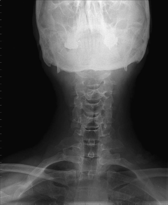
76 A 50-year-old man was troubled by a recent onset of arm pain and numbness. He had no neurologic deficit. Mild cervical spondylosis was seen on magnetic resonance imaging (MRI). An anteroposterior (AP) C-spine x-ray is remarkable for the presence of a cervical rib (Fig. 76-1). Thoracic outlet syndrome A course of conservative therapy with pain control and physical therapy was initiated, and the patient will make follow-up visits to the clinic. Thoracic outlet syndrome is a controversial diagnosis that refers to the myriad of symptoms caused by compression of the brachial plexus, subclavian vein, or sub-clavian artery. Brachial plexus compression (C8 and T1) occurs most frequently and predominantly in females. In addition to medial arm and fourth and fifth digit pain, cold intolerance, headache, and fine motor weakness can occur with neurologic compression. Venous compression manifests as a painful, edematous arm, and arterial compression causes claudication, arm pallor, and pain. In addition, the latter patient will have lower blood pressure in the affected arm. Often the physical examination is normal. Most provocative tests, like Adson’s maneuver, are not very reliable, although asking the patient to abduct his arms 90 degrees from the thorax, flex his elbows, and rapidly open and close his hands for 3 minutes can reproduce thoracic outlet symptoms. On radiologic imaging, a cervical rib, elongation of the C7 transverse process, or a congenital fibromuscular band may be noted. The anterior and middle scalene muscles may also be the source of compression. There is some evidence that repetitive activity may play a part in this syndrome.
Cervical Rib
Presentation
Radiologic Findings
Diagnosis
Treatment
Discussion

Cervical Rib
Only gold members can continue reading. Log In or Register to continue

Full access? Get Clinical Tree


