Circuits and Cells
THE NEURONAL CELL
The human brain is the most complex organ known to exist in the universe. Its weight is just 3% of the body, but it consumes 17% of the body’s energy. The workhorse of the brain is the neuron. It is estimated that we have 100 billion neurons with 100 trillion connections. When we think of a neuron, we are typically thinking of a pyramidal neuron in the cerebral cortex. These neurons have a diamond-shaped cell body and usually reside in layers III or V of the gray matter (Figure 3.1).
Let us start with a brief review of cell biology. The cell body of the neuron is full of the usual assortment of organelles, although not in the same proportions as seen in non-neural cells. Structures such as the endoplasmic reticulum (ER) and mitochondria are found more frequently in neurons than in other brain cells, presumably because of the increased need for protein synthesis and energy production. The instructions for the functioning of the cell are contained in the DNA, which resides in the nucleus. The instructions are read when the DNA is transcribed into messenger ribonucleic acid (mRNA), which is translated into proteins in the cytoplasm (Figure 3.2). As mentioned in Chapter 1, this process is often called gene expression— two of our favorite words.
The ribosome is the organelle in which mRNA is translated into proteins (Figure 3.3). The ribosomes are usually attached to the rough ER, but can also be floating freely in the cytoplasm. The proteins, once they are refined, are used by the cell for structural (e.g., receptors), functional (e.g., enzymes), or communication (e.g., neuropeptides) purposes, to name a few.
The Golgi apparatus, which looks like ER without the ribosomes, is where much of the “posttranslation” refinement, sorting, and storage of proteins occurs. This structure enables proteins to be appropriately transported to distant sites within the cell.
The mitochondria are the remarkably abundant energy generators of the neuron. The brain requires considerable energy, even at rest, just to maintain an electrical gradient poised to respond at a moment’s notice. The mitochondria convert adenosine diphosphate into adenosine triphosphate (ATP) and it is ATP that the cell uses to perform its functions.
The dendrites are the part of the neuron that sprout off the cell body and look like tree branches. They are often called the ears of the neuron, because they receive input from other neurons and relay the signal to the cell body. Most dendrites have “little knobs” along their stalks that are called dendritic spines. Each spine is the postsynaptic receptor for an incoming signal from another neuron (Figure 3.4).
The morphology (structure and density) of the dendritic spines has been a source of considerable interest since Ramón y Cajal first identified them over 100 years ago. Spines can change in shape, volume, and number with remarkable speed and frequency. This plasticity is one of the great discoveries of modern neuroscience. We mention spines throughout this book as their morphology changes in a number of conditions, including substance abuse, mental retardation, schizophrenia, and learning (see Point of Interest).
The axon is perhaps the most unique structure of the neuron. Starting at the axon hillock and running anywhere from a few micrometers to the entire length of the spinal cord, the axon can transmit a signal quickly without degradation to other
neurons or end organs. For this reason, the axon is often conceptualized as the telephone wire of the brain. Because the axon is devoid of ribosomes and incapable of protein synthesis, a process called axoplasmic transport enables the neuron to send material down the microtubules to the distal ends of the cell.
neurons or end organs. For this reason, the axon is often conceptualized as the telephone wire of the brain. Because the axon is devoid of ribosomes and incapable of protein synthesis, a process called axoplasmic transport enables the neuron to send material down the microtubules to the distal ends of the cell.
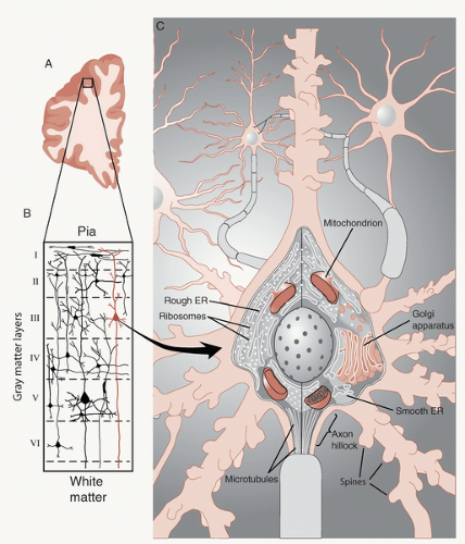 FIGURE 3.1 • A. Cross section of the right prefrontal cortex (PFC). B. The six layers of neurons in the gray matter of the PFC. C. A stereotypical pyramidal neuron found in layer III of the cerebral cortex. ER, endoplasmic reticulum. (Adapted from Bear MF, Connors BW, Paradiso MA, eds. Neuroscience: Exploring the Brain. 3rd ed. Baltimore, MD: Lippincott Williams & Wilkins; 2007.) |
The terminal end of the axon forms the synapse (Figure 3.5). This is where one neuron talks to another—if the dendrites are the ears, the synapse is the mouth. Here the electrical signal streaming down the axon is converted into a chemical signal, so the impulse can pass from one cell to another. The neurotransmitters that form the basis of the chemical signal are stored in vesicles. When released, they diffuse across the synaptic cleft to receptors on the postsynaptic dendrite (more on this later in the chapter).
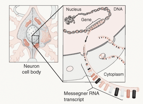 FIGURE 3.2 • Messenger ribonucleic acid (mRNA) carries the genetic instructions from the nucleus to the cytoplasm where translation into proteins occurs. (Adapted from Bear MF, Connors BW, Paradiso MA, eds. Neuroscience: Exploring the Brain. 3rd ed. Baltimore, MD: Lippincott Williams & Wilkins; 2007.) |
ELECTRICAL SIGNALING
All living cells maintain a negative internal electronic charge relative to the fluid outside of the cell—roughly -60 mV in a neuron. Nerve cells use the depolarization (rapid change in the electrical charge) to communicate with other nerves or end organs. There are two basic steps in this process. The neuron first receives signals through the dendrites, which are called postsynaptic potentials. Second, the cell sums the incoming impulses and if they are high enough then it sends an impulse down the axon, which is called an action potential.
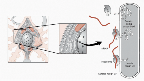 FIGURE 3.3 • Messenger ribonucleic acid (mRNA) binds to a ribosome, initiating protein synthesis. Proteins synthesized on the rough endoplasmic reticulum (ER) as shown are eventually inserted into the membrane. Proteins synthesized on free ribosomes (not shown) are utilized in the cytosol. (Adapted from Bear MF, Connors BW, Paradiso MA, eds. Neuroscience: Exploring the Brain. 3rd ed. Baltimore, MD: Lippincott Williams & Wilkins; 2007.) |
POINT OF INTEREST
Enhanced branching and spine formation have been found consistently in rats raised in enriched environments when compared with rats raised in standard wire cages. The neurons in the figure below are from rats raised in different environments. Note the increased branching (also called arborization) of the neuron from the rat raised in the enriched environment. Abundant dendritic branching with multiple connections seems to be a microscopic sign of a healthy active brain.
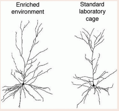 From Kolb B, Forgie M, Gibb R, et al. Age, experience and the changing brain. Neurosci Biobehav Rev. 1998;22(2):143-159. |
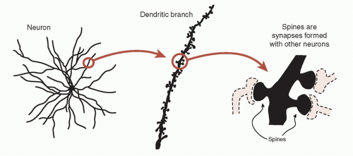 FIGURE 3.4 • Spines are the “little knobs” on the dendrites. They are on the receiving side of the electrochemical signal from other neurons—also called the postsynaptic membrane. |
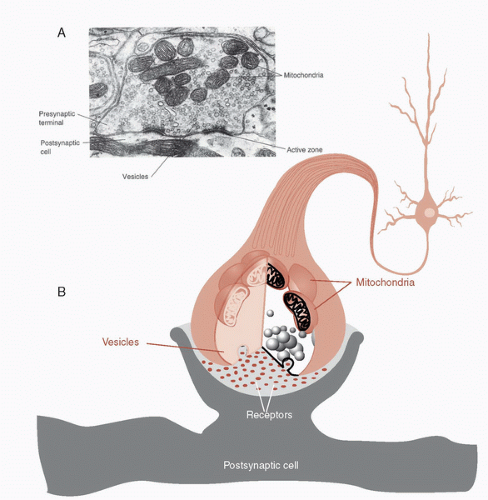 FIGURE 3.5 • A synapse as seen with an electron microscope (a) and in a schematic drawing (B). Note the high concentration of vesicles filled with neurotransmitter and mitochondria to power the rapid processing. (Adapted from Bear MF, Connors BW, Paradiso MA, eds. Neuroscience: Exploring the Brain. 3rd ed. Baltimore, MD: Lippincott Williams & Wilkins; 2007.) |
Postsynaptic Potentials
A single pyramidal cell will receive input from 1 to 100,000 neurons through the postsynaptic synapses (spines) on the dendrites and cell body. When the neurotransmitters bind with the receptor at the postsynaptic synapse, ions flow into the neuron and change the electrical potential making it more positive or more negative—or what is called depolarization and hyperpolarization.


Stay updated, free articles. Join our Telegram channel

Full access? Get Clinical Tree


