Cistern, Subarachnoid Space Normal Variants
Karen L. Salzman, MD
DIFFERENTIAL DIAGNOSIS
Common
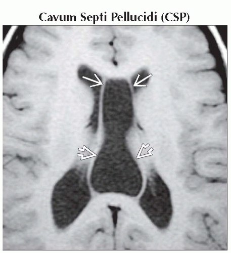
Cavum Septi Pellucidi (CSP)
Mega Cisterna Magna
MR Artifacts, Flow-Related
Enlarged Subarachnoid Spaces
Less Common
Cavum Velum Interpositum (CVI)
Enlarged Optic Nerve Sheath
Rare but Important
Blake Pouch Cyst
Liliequist Membrane
ESSENTIAL INFORMATION
Key Differential Diagnosis Issues
Normal variants have CSF density/intensity
Important to recognize normal variants & not mistake for more ominous pathology
Helpful Clues for Common Diagnoses
Cavum Septi Pellucidi (CSP)
Elongated finger-shaped CSF collection between frontal horns of lateral ventricles
Posterior continuation between fornices often associated (cavum vergae)
Mega Cisterna Magna
Enlarged cisterna magna communicates freely with 4th ventricle & basal cisterns
Large posterior fossa
Normal vermis
Cistern crossed by falx cerebelli, tiny veins
Occipital bone may appear scalloped
MR Artifacts, Flow-Related
CSF flow artifact is common in basal cisterns, ventricles
Commonly seen on FLAIR MR
Artifact often extends outside skull
Enlarged Subarachnoid Spaces
Idiopathic enlargement of subarachnoid spaces (SAS) during first year of life
Increased head circumference (> 95%)
Resolves without therapy by 12-24 months
Helpful Clues for Less Common Diagnoses
Cavum Velum Interpositum (CVI)
Triangular-shaped CSF space between bodies of lateral ventricles, below fornices, above 3rd ventricle
Often elevates, splays fornices & causes inferior displacement of internal cerebral veins & 3rd ventricle
Enlarged Optic Nerve Sheath
May occur as normal variant
Occurs in idiopathic intracranial hypertension (pseudotumor cerebri), NF1
Helpful Clues for Rare Diagnoses
Blake Pouch Cyst
Failure of regression of Blake pouch cyst causes compression of basal cisterns
Free communication of 4th ventricle with prominent inferior CSF space
Liliequist Membrane
Thin arachnoid membrane separates suprasellar, interpeduncular, & prepontine cisterns
Image Gallery
 Axial T1WI MR shows a cavum septi pellucidi
 with posterior extension into a cavum vergae with posterior extension into a cavum vergae  , seen as a CSF-signal collection that lies between the bodies of the lateral ventricles. , seen as a CSF-signal collection that lies between the bodies of the lateral ventricles.Related posts:Stay updated, free articles. Join our Telegram channel
Full access? Get Clinical Tree
 Get Clinical Tree app for offline access

|