Figure 5.1
Plain radiograph of the lumbar spine, lateral projection. There is a chronic fracture of the second lumbar vertebral body (arrow). The vertebra has been compressed and has lost significant height with mild forward angulation of the spinal column (kyphosis) at this level
What Does the Procedure Entail?
The patient is placed in either the standing or lying position. Then an x-ray tube is positioned over the relevant body part and a quick snapshot is taken. Multiple views may be needed. This painless procedure is over quickly.
Computed Tomography (CT)
What Is It?
CT is a sophisticated piece of x-ray based equipment that obtains a cross-sectional visualization of the body. It plays a central role in the workup and management of neurological disease. Similar to plain radiography, CT uses x-rays that are carefully controlled and monitored. A rotating x-ray tube and detector array combines with a complex computer to create images that represent those structures being imaged. The images produced are sequential layers or slices of the area of interest. The scans are quick to perform in most cases.
What Does the Procedure Entail?
The patient lies on a table that moves part or all of the body into the center of the scanner, which looks like a giant doughnut. Patients are sometimes given a special contrast agent into a vein, depending on the needs of the study.
CT of the Brain
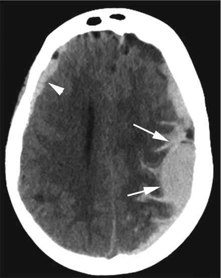
The most common use of CT in neuroradiology is imaging of the brain. CT is particularly helpful in detecting acute intracranial hemorrhage (bleeding), contusions (bruising), stroke, edema (swelling) and hydrocephalus (enlargement of the normal spaces that contain cerebrospinal fluid). CT can also be useful in evaluating certain brain tumors, particularly in patients who cannot have an MRI (Fig. 5.2).


Figure 5.2
CT of the brain. There is acute hemorrhage (white) overlying the left (arrows) and to a lesser extent the right (arrowhead) sides of the brain. This patient was involved in a high velocity motor vehicle accident. The head CT, performed shortly after arrival in the emergency room, facilitated rapid diagnosis and subsequent emergent life-saving neurosurgical evacuation of the larger left-sided brain hemorrhage
CT Angiography (CTA)
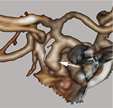
CT angiography is the term used to describe the imaging of arteries (CT arteriography) and/or veins (CT venography). CTA enables neuroradiologists to view the blood vessels of the head without the surrounding brain tissue or skull. First, a patient is given a contrast agent that makes the blood vessels visible. A CT scan is then performed with sub-millimeter thin images. Finally, computer modeling is used to see the blood vessels. This method has revolutionized the diagnosis of ruptured brain aneurysms and other abnormalities of blood vessels such as arteriovenous malformations (AVMs) (Fig. 5.3).


Figure 5.3
CT angiography of the brain with 3D reconstruction image. A patient admitted with sudden acute life-threatening brain hemorrhage was found to have a ruptured 7 mm brain aneurysm arising from the distal right carotid artery (arrow). The patient was brought to the operating room that night for aneurysm repair and made a full recovery
CT Perfusion (CTP)
This technique measures blood flow to the brain and offers an indirect assessment of blood volume, flow, and transit through the region of imaged brain. A contrast agent is administered and the brain is rapidly imaged to understand how blood moves through brain tissue. It can help characterize certain brain tumors but is more commonly used in the assessment of acute stroke by identifying damaged but still salvageable portions of the brain.
CT of the Spine
CT scans of the spine are typically done in segments of the body: cervical (neck region), thoracic (chest region), or lumbar (abdominal region). Cross-sectional images are viewed separately and can be combined or reformatted with others in different planes to see alignment of the spine. Because of its strength in the assessment of bony structures compared with soft tissues such as ligaments and muscles, CT is the gold standard in the initial imaging of serious spinal trauma. It also provides excellent evaluation of degenerative changes of arthritis and positioning of surgically placed metal screws and cages following spine surgery.
CT-guided Intervention
CT scans can be performed prior to surgery or during a procedure to improve localizing an area of interest. If the scan is going to be used during surgery, the data is sent to the operating room and used for operative planning. Like a GPS roadmap, the surgeon can view the images and use that data to guide his/her instruments with more precision. Another helpful use of CT is to assist the placement of small needles used for biopsies to obtain tissue or injection of steroid in the management of chronic back pain with or without radiculopathy, the medical term for what is commonly known as “sciatica”. The CT-guided images allow for high accuracy during these types of procedures.
Magnetic Resonance Imaging (MRI)
What Is It?
MRI has been widely used since the 1980s and has significantly improved since that time. MRI is particularly useful in imaging disorders of the brain and spinal cord. MRI works by positioning the patient in a very strong magnetic field and then using radio waves which are received by detectors. Complex computer analysis then determines the different anatomic and chemical structures of the tissues being imaged. These result in exquisite detail with better resolution of soft tissues, including the brain, compared to any other radiological modality. One of the advantages is that MRI does not use ionizing radiation. A disadvantage is that MRI may be contraindicated in individuals with certain implantable metallic devices, including most cardiac pacemakers and neurostimulators. If you have metal in your body, it is important to tell the MR technologist before entering the magnet area.
Stay updated, free articles. Join our Telegram channel

Full access? Get Clinical Tree


