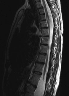
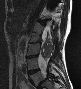
81 A 41-year-old woman with known congenital abnormalities of the spine complained of lower extremity radiculopathy. She had decreased left S1 sensation and a tuft of hair overlaying the thoracolumbar junction. Magnetic resonance imaging (MRI) of the whole spine was reviewed, and it is notable for numerous pathologies. In addition to segmentation anomalies and spondylolysis, there are several intradural findings. The T2-weighted sagittal MRI of the thoracic spine shows intramedullary hyperintensity (Fig. 81-1). The same sequence shows a scoliotic lumbar spine, with grade II L5-S1 spondylolis-thesis and a large heterogenous lesion displacing the cauda equina (Fig. 81-2). There is also diastematomyelia type II (Figs. 81-3 and 81-4) that extends from the lumbar to thoracic spine. The diastematomyelia tapers into a common thecal sac above and below the spur, as noted in the myelogram (Fig. 81-5). FIGURE 81-1 MRI showing a thoracic intramedullary lesion with a syrinx. Spina bifida occulta, with diastematomyelia, tethered cord, teratoma, and spondylolisthesis
Congenital Abnormality of the Spine
Presentation
Radiologic Findings

Diagnosis

Congenital Abnormality of the Spine
Only gold members can continue reading. Log In or Register to continue

Full access? Get Clinical Tree


