(1)
Marina Spine Center, Marina del Rey, CA, USA
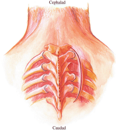
The costotransversectomy is a thoracic spine approach that can be made in one of two positions: prone [1 , 2] or in the lateral decubitus position tilted anteriorly 20 degrees [3]. Either of two skin incisions, midline or paraspinous, and one of three fascial incisions, midline, longitudinal paraspinous or transverse paraspinous, can be used. The pathology determines the exact approach. For biopsy and exposure of an intervertebral disc or vertebral body, position the patient prone, make a midline incision, and retract the paraspinous muscle mass laterally (Fig. 44.1). A transverse incision in the paraspinous fascia and muscle may be necessaiy. For decompression after Harrington rod insertion, a long midline incision allows adequate lateral retraction to expose the costotransverse joint. Resect the transverse process, resect the pedicle, and decompress the spinal canal.4Adequate interbody fusion can be most difficult with the prone position, midline incision approach. Optimum exposure for resection of an entire vertebral body and two intervertebral discs followed by a strut graft is through a semilateral decubitus position, a paraspinous skin incision, and a paraspinous fascial incision [3].


Fig. 44.1
Costotransversectomy approach. Position the patient prone. Make a midline incision and retract the paraspinous muscle mass laterally. A transverse incision of the paraspinous fascia and muscle may be needed. Make the incision of sufficient length to allow this lateral retraction. Alternatively, a curvilinear skin incision, its midportion 6 cm from the midline and the end approximately 3 cm from the midline, may be used to expose directly the lateral border of the paraspinous musculature. For the more lateral incision, the paraspinous musculature is dissected from lateral to medial toward the midline. For the midline incision, the paraspinous musculature is elevated from the posterior elements medial to lateral
1.
Position the patient prone on the operating table on a suitable frame. The horseshoe-shaped cushion with two chest pads or a four-poster operating frame made of radiolucent material allows excellent chest excursion and support during the operation. For approaches in the upper thoracic spine, the patient’s arms are at the side and the head should be carefully positioned on the well-padded cervical headrest with carehll padding of the forehead and malar eminences. Pay particular attention to the eyes and allow no pressure on them. Securely anchor the endotracheal tube. For approaches to the lower thoracic spine, position the arms at 90 degrees to the chest, the elbows well padded such as for a lumbar operation. The patient’s head can be turned.
2.
After prepping and draping, make a midline incision of sufficient length to allow retraction of paraspinous musculature for work lateral to the costotransverse joint (Fig. 44.1). Alternately, a curvilinear skin incision with its midportion 6 cm from the midline and the two ends of the incision approximately 3 cm from the midline can be used to expose directly the lateral outer border of the paraspinous musculature.
3.
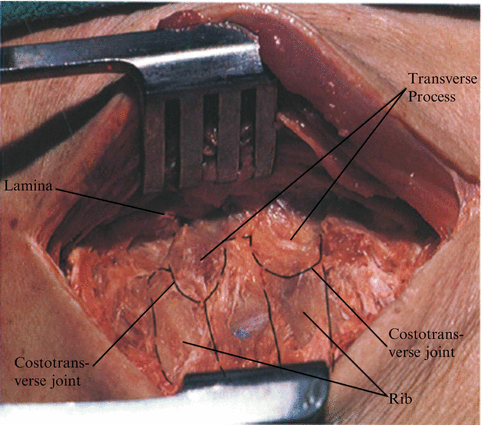
Extend the midline incision as in any posterior spinal dissection, cutting with cautery to the spinous processes and removing muscle attachments from the lamina with a sharp periosteal elevator (Fig. 44.2). Continue this subperiosteal removal of musculature and fascia laterally onto the rib beyond the costovertebral joint. For unilateral retraction use the single-tooth self-retaining lamina retractor, with the single-tooth edge in the spinous process and the wide retractor blade beneath the paraspinous musculature. The paraspinous incision cuts through the thoracolumbar fascia laterally and elewtes the paraspinous muscle mass medially.


Fig. 44.2
With sharp dissection, remove the periosteum from the rib and the capsule of the costotransverse process. Open the costotransverse joint
4.


With sharp periosteal dissection, remove the periosteum from the rib, the capsule of the costotransverse joint, and the muscle and fascial tissue covering of the transverse process (Fig. 44.2).
Stay updated, free articles. Join our Telegram channel

Full access? Get Clinical Tree


