Coverings of the central nervous system
The CNS is supported and protected by bone and membranous coverings. The brain is located within the cranial cavity of the skull and the spinal cord lies in the vertebral, or spinal, canal within the vertebral column, or spine. Within their bony coverings, the brain and spinal cord are invested by three concentric membranous envelopes. The outermost membrane is the dura mater, the middle layer is the arachnoid mater and the innermost layer is the pia mater. The vertebral column and spinal meninges are described in Chapter 8; consequently, only the skull and cranial meninges are considered here.
Skull
The brain lies on the floor of the cranial cavity, which, together with the bones of the cranial vault, provides support and protection from physical injury. The floor of the cranial cavity consists of three fossae. Each of these accommodates particular parts of the brain and possesses foramina through which cranial nerves and blood vessels enter and leave the cranial cavity (Fig. 5.1).
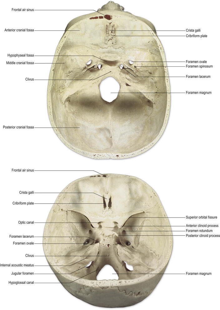
Anterior cranial fossa
The anterior cranial fossa is formed by the frontal, ethmoid and sphenoid bones. It contains the frontal lobes of the brain. The greater part of the floor of the anterior cranial fossa consists of the frontal bone and it also forms the roof of the orbit. The part of the frontal bone that forms the anterior wall of the fossa contains the frontal air sinus. In the midline of the floor of the anterior cranial fossa is the ethmoid bone. In the midline, a sharp ridge, the ‘crista galli’, is the anterior point of attachment of the dural falx cerebri. In an elongated depression on either side of the crista galli lie the cribriform plates of the ethmoid bone. These accommodate the olfactory bulbs. The bone of the cribriform plate is peppered with small perforations through which the fascicles of the olfactory nerve enter the cranial cavity from the nasal cavity to attach to the olfactory bulb.
Middle cranial fossa
The middle cranial fossa is formed by the sphenoid and temporal bones. In the midline, the body of the sphenoid forms a deep depression, the hypophyseal fossa, encompassed by four spurs of bone, the anterior and posterior clinoid processes. In the hypophyseal fossa lies the hypophysis, or pituitary gland. Lateral to the body of the sphenoid, the rest of the middle cranial fossa contains the temporal lobes of the cerebral hemisphere. The middle cranial fossa contains numerous points of entry to, and exit from, the cranial cavity for cranial nerves and blood vessels. In particular:
Posterior cranial fossa
The posterior cranial fossa is formed by the occipital and petrous temporal bones. Anteriorly, in the midline, it forms a steep, smooth slope (the clivus) that is continuous with the body of the sphenoid bone, posterior to the hypophyseal fossa. The brainstem rests upon the clivus, the medulla passing through the foramen magnum to become continuous with the spinal cord. The foramen magnum also admits the vertebral arteries and the spinal root of the accessory (XI) nerve. In the lateral wall of the foramen magnum lies the hypoglossal canal through which the hypoglossal (XII) nerve leaves the cranial cavity. Between the occipital and petrous temporal bones lies the large jugular foramen through which pass the internal jugular vein, and the glossopharyngeal (IX), vagus (X) and accessory (XI) nerves. In the vertical wall of the petrous temporal bone is located the internal auditory (acoustic) meatus, which transmits the vestibulocochlear (VIII) and facial (VII) nerves. The cerebellum rests on the floor of the posterior cranial fossa.
 Raised intracranial pressure
A space-occupying lesion is an expanding focal lesion such as a tumour, haematoma or abscess. Since the cranial cavity is closed and unyielding, the brain is distorted and displaced downwards, towards the foramen magnum, as the intracranial pressure rises (Fig. 5.2). The patient complains of headache, vomiting, blurring of vision and drowsiness. The optic discs are swollen (papilloedema), signs of brainstem dysfunction are found, and coma and death supervene if the pressure is not relieved by neurosurgery (craniotomy). Benign intracranial hypertension is caused by generalised swelling of the brain in the absence of a focal space-occupying lesion. It often occurs in obese young women; the syndrome of raised intracranial pressure mimics a brain tumour, hence the old designation, ‘pseudo tumour cerebri’.

< div class='tao-gold-member'>
Related posts:


Stay updated, free articles. Join our Telegram channel

Full access? Get Clinical Tree


