Cranial Nerves
The Eye 1 – Pupils, Acuity, Fields
BACKGROUND
Examination of the eye can provide very many important diagnostic clues for both general medical and neurological diseases.
Examination can be divided into:
3 Acuity
Abnormalities may arise from:
It is essential to test acuity with the patient’s correct glasses.
4 Fields
The organisation of the visual pathways means different patterns of visual field abnormality arise from lesions at different sites. The normal visual pathways are given in Figure 7.1.
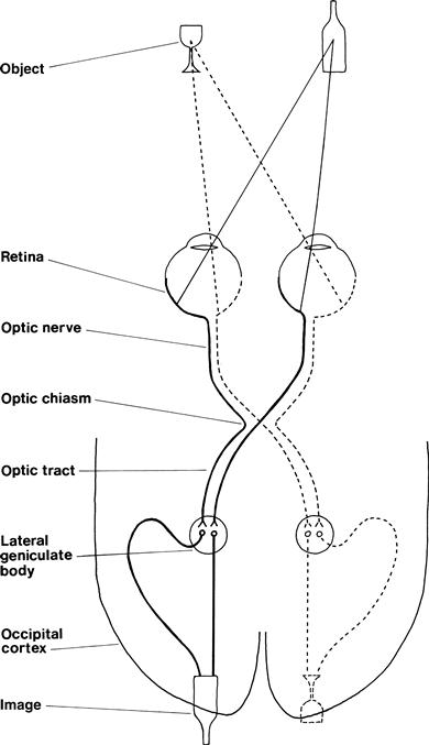
The visual fields are divided vertically through the point of fixation into the temporal and nasal fields. Something on your right as you look ahead is in the temporal field of your right eye and the nasal field of your left eye.
The visual fields are described from the patient’s point of view.
Field defects are said to be homonymous if the same part of the visual field is affected in both eyes. This can be congruous (the field defects in both eyes match exactly) or incongruous (the field defects do not match exactly).
Testing the fields is very useful in localisation of a lesion (Table 7.1).
Table 7.1
| Type of defect | Site of lesion |
| Monocular field defect | Anterior to optic chiasm |
| Bitemporal field defect | At the optic chiasm |
| Homonymous field defect | Behind the optic chiasm |
| Congruous homonymous field defect | Behind the lateral geniculate bodies |
The normal visual fields for different types of stimuli are very different. The normal field for moving objects or large objects is wider than for objects held still or small objects. The normal field for recognition of coloured objects is more limited than for monochrome. It is useful to test this on yourself. Look straight into the distance in front of you and put your hands out straight to your side. Wiggle your fingers and, keeping your arms straight, gradually bring your arms forward until you can see your moving fingers. Repeat this holding a small white object, and then with a red object until you can see that it is red. You will appreciate the different normal fields for these different stimuli.
1 GENERAL
WHAT TO DO
Look at the patient’s eyes and note any difference between the two sides.
Look at the level of the eyelid; particularly note asymmetry.
Look at the position of the eye.
Beware the false eye—usually obvious on closer inspection.
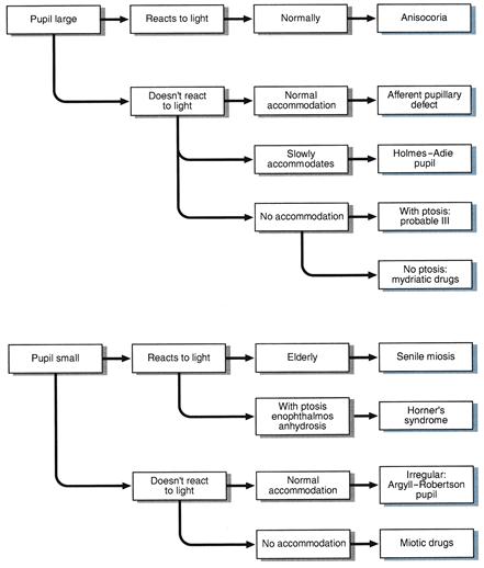
2 PUPILS
WHAT TO DO IN A CONSCIOUS PATIENT
(For pupillary changes in an unconscious patient, see Chapter 27.)
Look at the pupils.
• Are they regular in outline?
• Are there any holes in the iris or foreign bodies (e.g. lens implants) in the anterior chamber?
Shine a bright light in one eye.
Place your finger 10 cm in front of the patient’s nose. Ask the patient to look into the distance and then at your finger.
Look at the pupils for their reaction to accommodation.
FURTHER TESTING
Swinging light test
What to do
Shine a bright light into one eye and then the other at about 1-second intervals. Swing the light repeatedly between the two. Observe the pupillary response as the light is shone into the eye.
Related posts:

Stay updated, free articles. Join our Telegram channel

Full access? Get Clinical Tree


