Critical Illness Polyneuropathy and Myopathy

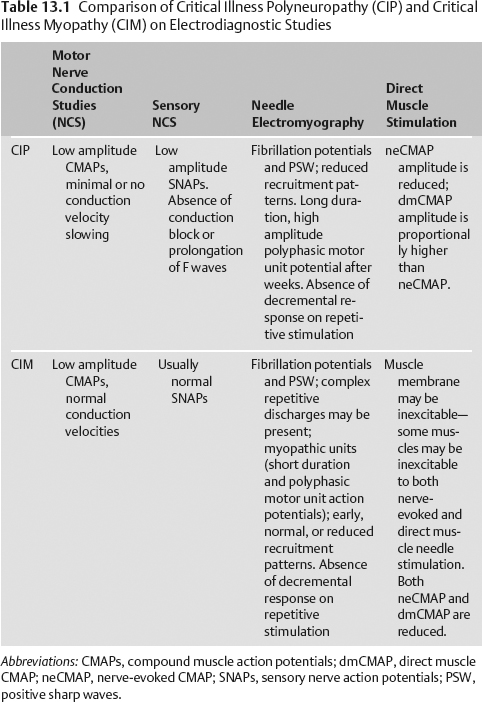
13 Critical Illness Polyneuropathy and Myopathy


Valerie Dechant, Eduardo Adonias de Sousa, and Kiwon Lee


Critically ill patients are at risk for developing severe weakness secondary to critical illness polyneuropathy (CIP) and/or critical illness myopathy (CIM). Weakness may progress to severe quadriparesis and muscle wasting.1 The incidence of CIP/CIM in critically ill patients has been reported as 33 to 44% of patients with prolonged admission to critical care settings. The incidence increases to nearly 70% when considering only patients with sepsis.15 Risk factors that have been suggested for CIP and CIM include:


  • Sepsis/systemic inflammatory response syndrome (SIRS):2,3,5,6 Sepsis/SIRS are proinflammatory states that result in the release of cytokines as well as microcirculatory changes. These inflammatory changes can result in CIP by direct injury to the nerve as well as increased permeability of the endoneurium to neurotoxins. The activation of proteosomes in the proinflammatory state can lead directly to muscle protein breakdown, leading to CIM. Elevations in proteosomes have been detected as early as 1 week into intensive care unit (ICU) treatment.7 Nerve conductions studies done as early as 3 days post-sepsis onset have shown findings consistent with CIP 2,4,6,8
  • Neuromuscular blocking agents (NMBAs): The role of NMBAs as a risk factor for CIM has been debated. Some authors suggest that the use of NMBAs, particularly aminosteroidal (vecuronium, pancuronium, rocuronium) NMBAs or NMBAs used in conjunction with corticosteroids or aminoglycosides increase the risk of CIM.9 These agents have been found to cause pharmacological muscle denervation, resulting in upregulation of acetylcholine receptors and glucocorticoid receptors in muscle.5,6
  • Corticosteroids: The risk of developing CIM/CIP from steroid use is dose dependent with higher doses and longer uses. High-dose steroids contribute to muscle wasting by decreasing myosin synthesis and increasing muscle turnover, as well as triggering skeletal muscle apoptosis. Patients with sepsis may be at a higher risk of steroid-induced CIM, as glucocorticoid receptors of muscle may be unregulated in the septic state.6,7
  • Hyperglycemia: Elevated blood sugars lead to insulin resistance that can prevent glucose uptake in skeletal muscle, leading to a depletion of energy stores. Tight control of blood sugars has been shown to reduce the risk of CIP.10


Case Example


A 57-year-old woman was initially admitted to the ICU with a chronic obstructive pulmonary disease (COPD) exacerbation. Despite treatment with prednisone and β agonist nebulizers, she subsequently developed respiratory failure and became ventilator dependent. Her hospital course was complicated by sepsis. It became difficult to wean the patient from the ventilator. On morning rounds she was noted to have decreased motor function in withdrawing from painful stimuli in all four extremities.


Questions


  • What was the dose and duration of steroid use?
  • Has the patient been persistently hyperglycemic?
  • Did the patient receive neuromuscular blockers, and if so, for how long?
  • What antibiotics did the patient receive?
  • Does the patient have any known neurologic history that may account for the weakness (i.e., underlying peripheral neuropathy)?

Urgent Orders


  • Discontinue neuromuscular blockade, steroids, and aminoglycosides if possible.

History and Examination


History


Physical Examination


Assess for symmetry in the exam: gross asymmetry (hemiparesis, paraparesis, hemisensory loss or crossed signs) suggests an etiology other than CIP/CIM. Prominent mental status changes, cranial nerve involvement, pain, bowel or bladder involvement, cerebellar signs or a sensory level should prompt further work-up.


Neurologic Examination


  • A full neurologic examination, including assessment of mental status, cranial nerves, motor skills, and reflexes, as well as a sensory and cerebellar exam, should be performed on all patients.
  • CIM: Mental status not affected; flaccid quadriparesis proximal > distal; muscle atrophy; facial weakness is common, but extraocular muscle weakness is uncommon; reflexes normal; sensation intact
  • CIP: Mental status not affected, preservation of cranial nerve function, limb weakness, atrophy, fasciculations possible, reduced or absent reflexes, length dependent loss of sensation (test with painful stimuli in less responsive/nonverbal patients)

Differential Diagnosis


  1. CIP/CIM
  2. Guillain-Barré syndrome (GBS): GBS is an acute demyelinating polyneuropathy that typically begins as an ascending paralysis accompanied by areflexia and sometimes follows a diarrheal illness. Coincidental occurrence of GBS in a critically ill patient is relatively rare. However, if the history is suspicious, a lumbar puncture can be done (best yield is 1 to 2 weeks from symptom onset). Elevated protein without elevation in leukocytes would be suspicious for GBS. Cerebrospinal fluid (CSF) studies should be normal in CIP/CIM. Nerve conduction studies in GBS should be consistent with a demyelinating neuropathy in the AIDP (acute inflammatory demyelinating polyneuropathy) subtype (i.e., severely prolonged distal motor latencies, severe motor conduction velocity slowing, severely prolonged F waves), often with an acquired demyelinating component (i.e., partial conduction blocks and abnormal temporal dispersion). The axonal variant of GBS seems to be less frequent in the Western world and more frequent in China and Japan.
  3. Cachetic myopathy: Critically ill patients can develop a subacute myopathy due to protein catabolism and disuse. Patients develop weakness and muscle atrophy. Type II muscle atrophy is seen histologically.
  4. Spinal cord lesions: A lesion to the cervical spinal cord can result in tetraparesis and should be considered if the clinical setting is appropriate. Lesions in the cord can initially cause flaccid paresis with decreased reflexes, followed subacutely by hyperreflexia and increased tone. A sensory level may be evident in a patient who is able to participate in a sensory examination. If the history or examination is suspicious, a magnetic resonance imaging (MRI) scan of the cervical cord with and without contrast may be appropriate.
  5. Underlying neuropathy-Toxins and medication effects: Often medications used in the ICU (i.e., neuromuscular blocking agents) can result in prolonged weakness and sedation. A nerve conduction study may help in this case. For patients who have been treated with neuromuscular blocking agents, “a train of four” (slow repetitive stimulation at 2 to 3 Hz at the median or ulnar nerve) may be used to determine if the medication’s effect is persisting.

Life-Threatening Diagnoses Not to Miss


  • Spinal cord lesion

Diagnostic Evaluation


Stay updated, free articles. Join our Telegram channel

Aug 30, 2016 | Posted by in NEUROSURGERY | Comments Off on Critical Illness Polyneuropathy and Myopathy

Full access? Get Clinical Tree

Get Clinical Tree app for offline access