Cystic Intrasellar Mass
Anne G. Osborn, MD, FACR
DIFFERENTIAL DIAGNOSIS
Common
Empty Sella (ES)
Intracranial Hypertension, Idiopathic
Less Common
Obstructive Hydrocephalus
Rathke Cleft Cyst
Craniopharyngioma
Arachnoid Cyst (AC)
Epidermoid Cyst
Neurocysticercosis Cyst
Rare but Important
Pituitary Apoplexy
Saccular Aneurysm (Thrombosed)
ESSENTIAL INFORMATION
Key Differential Diagnosis Issues
Cystic mass originating WITHIN sella vs. intrasellar extension from suprasellar lesion
Intrasellar extension of suprasellar lesion > cystic intrasellar mass
Helpful Clues for Common Diagnoses
Empty Sella (ES)
Small crescent of compressed pituitary gland lines bottom of sella turcica
“Primary” ES considered normal variant
“Secondary” = surgery, pituitary infarction
Intracranial Hypertension, Idiopathic
“Pseudotumor cerebri” F > > M
Empty sella ± dilated optic nerve sheaths, small ventricles
Helpful Clues for Less Common Diagnoses
Obstructive Hydrocephalus
Anterior recesses of 3rd ventricle enlarge
Herniate inferiorly into sella
If chronic may expand, erode bony sella
Rathke Cleft Cyst
Usually < 1 cm; can be giant, erode sella
45% have “intracystic nodule”
± “Claw sign” (enhancing rim of pituitary around nonenhancing cyst)
Craniopharyngioma
Truly intrasellar craniopharyngioma rare
If no Ca++ difficult to distinguish from Rathke cleft cyst
Arachnoid Cyst (AC)
Truly intrasellar AC rare
Usually extension from suprasellar AC
Epidermoid Cyst
Suprasellar location < off-midline
Neurocysticercosis Cyst
Suprasellar cysts → intrasellar
Helpful Clues for Rare Diagnoses
Pituitary Apoplexy
Can be life-threatening (secondary to pituitary insufficiency)
Acutely may present as necrotic, rim-enhancing mass
Saccular Aneurysm (Thrombosed)
Medially projecting from cavernous ICA
If thrombosed may appear low signal intensity on T1 C+ scans
Image Gallery
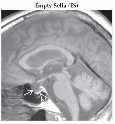 Sagittal T1WI MR shows empty sella with herniation of CSF through the diaphragma sellae
 flattening the pituitary gland inferiorly against the sellar floor flattening the pituitary gland inferiorly against the sellar floor  . .Related posts:Stay updated, free articles. Join our Telegram channel
Full access? Get Clinical Tree
 Get Clinical Tree app for offline access

|
