Fig. 19.1
Axial noncontrast head CT demonstrating a left parieto-occipital contrecoup contusion with surrounding hypodensity and subtle bifrontal hypodensities with 5 mm of left to right midline shift. There is a right frontal EVD in place

Fig. 19.2
Coronal head CT at the posterior border of the sphenoid sinus demonstrating a wide bifrontal craniectomy with extension to the middle cranial fossa floor

Fig. 19.3
Sagittal CT showing extension of the craniectomy posterior to the coronal suture
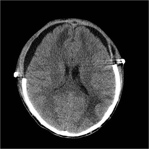
Fig. 19.4
Bilateral subdural hygromas that resolved spontaneously within 3 days
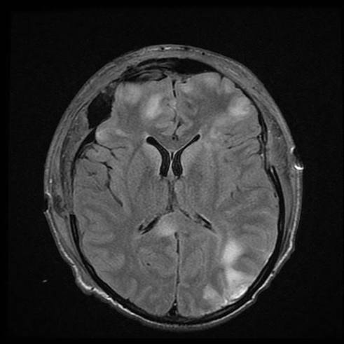
Fig. 19.5
T2 FLAIR sequence 1 month after accident showing signal intensity within the right parieto-occipital, bifrontal, and splenium consistent with diffuse axonal injury
A 21-year-old male was involved in a T-bone collision while riding a motorcycle wearing a full face helmet. He was a GCS 3T on arrival to the hospital. His head injuries involved a left parieto-occipital contrecoup contusion and subtle bifrontal hypodensities and multiple orthopedic injuries. An EVD was placed and his intracranial pressures were managed medically for the first 5 days. Eventually the right parieto-occipital contusion and bifrontal hypodensities evolved causing 5 mm of left to right midline shift and medically refractory elevated ICPs > 20 mmHg. A wide bifrontal craniectomy was performed. He developed bilateral subdural hygromas 2 weeks after surgery that resolved spontaneously after 3 days of observation. He underwent a bifrontal cranioplasty 4 months after the craniectomy. He was discharged to neuro-rehab with a GCS 14. He has moderate cognitive changes and emotional lability consistent with his frontal lobe injury.
Case 2: (see Figs. 19.6, 19.7 and 19.8)
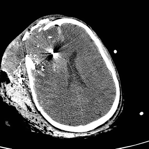

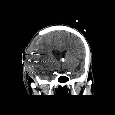
Fig. 19.6
Initial head CT demonstrating comminuted fractures of the frontal, temporal, and parietal bones, metallic fragmentation, traumatic subarachnoid hemorrhage around the anterior falx, and extensive soft tissue hemorrhage
Fig. 19.7
Right frontotemporoparietal craniectomy (11 cm × 14 cm) with a left frontal EVD in place. The posterior extension stopped at a fracture line within the parietal bone and further removal of bone was not thought to be necessary at the time. The craniectomy extended inferiorly to the floor of the middle cranial fossa with less than a centimeter of temporal squamosal bone remaining
Fig. 19.8
Right frontotemporoparietal craniectomy (11 cm × 14 cm) with a left frontal EVD in place. The posterior extension stopped at a fracture line within the parietal bone and further removal of bone was not thought to be necessary at the time. The craniectomy extended inferiorly to the floor of the middle cranial fossa with less than a centimeter of temporal squamosal bone remaining
A 27-year-old male deployed overseas suffered a gunshot wound to the head from an unknown caliber weapon. He underwent an emergent cricothyroidostomy and was transferred to higher level care with an admission GCS score of 8T. Initial head CT demonstrated significant right maxillary, orbital, frontal, and zygomatic fractures, retained metal fragmentation within the right frontal lobe and effacement of the frontal horn of the right lateral ventricle. A left frontal EVD was placed but within 24 h his ICPs were refractory to medical management. He underwent a right unilateral craniectomy in theater. He was transferred back to Walter Reed for further care and rehabilitation. One month after the accident he was discharged to neuro-rehab with a GCS 14 and moderate impairment in judgment and insight. He underwent cranioplasty 6 months after the injury.
Case 3: (see Figs. 19.9, 19.10, 19.11, 19.12, 19.13, 19.14 and 19.15)
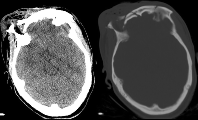
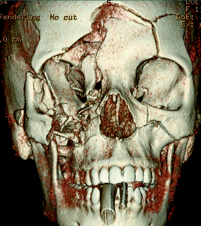
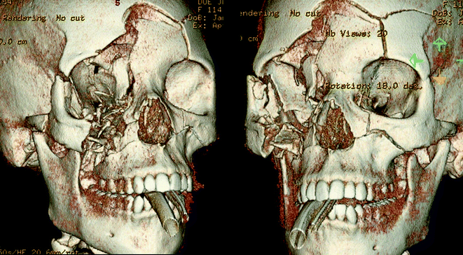

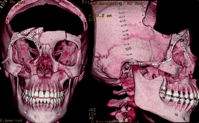
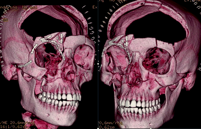
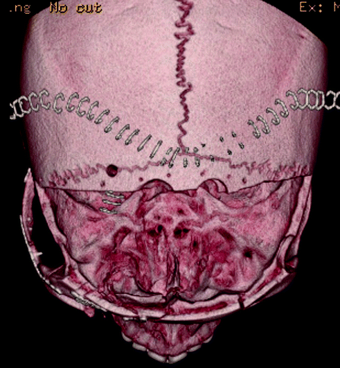
Fig. 19.9
Noncontrasted CT of head demonstrating comminuted fracture of the orbital bandeau and orbital roof and extra-cranial soft tissue swelling
Fig. 19.10
Three-dimensional reconstruction showing complex cranial, facial, and orbital fractures
Fig. 19.11
Three-dimensional reconstruction showing complex cranial, facial and orbital fractures
Fig. 19.12
Postoperative noncontrasted CT of head demonstrating blossoming bifrontal contusions
Fig. 19.13
Postoperative three-dimensional reconstruction demonstrating cranial decompression, reconstruction of zygomatic, maxillary, orbital bandeau, and orbital roof fractures
Fig. 19.14
Postoperative three-dimensional reconstruction demonstrating cranial decompression, reconstruction of zygomatic, maxillary, orbital bandeau, and orbital roof fractures
Fig. 19.15
Postoperative three-dimensional reconstruction demonstrating cranial decompression, reconstruction of zygomatic, maxillary, orbital bandeau, and orbital roof fractures
A 30-year-old female ejected from a motor vehicle after an unrestrained, head-on collision sustained direct craniofacial depression from a highway shoulder rail. On arrival, she was GCS 8T (E2-M5-V1T) and moving extremities symmetrically. She had gross brain matter and mixed serosanguinous fluid draining from an open facial laceration that extended from the middle of her forehead through her upper lip.
There are several intra-operative details that should be considered in conjunction with OMFS, Plastics, ENT and Oculoplastics teams to achieve several goals at the time of surgery: (1) decompression of cranial contents if there is a mass lesion or malignant edema, (2) primary repair of dural injuries using autograft, allograft, or dural substitutes, (3) reconstruction of anterior fossa floor and orbital bandeau, (4) thorough cranialization and exenteration of the frontal sinus, (5) reconstruction of severe, displaced facial and orbital injuries. A bicoronal incision was made and the subgaleal space was dissected anteriorly to the superior orbital rim, keeping the pericranium intact. A vascularized pericranial pedicle was incised beyond the incision posteriorly and up to the superior temporal line bilaterally; the pedicle was carefully dissected anteriorly to the superior orbital rim as well. This maneuver is critical for obtaining a large, vascularized autograft used for dural repair and reconstruction of the anterior fossa floor. A wide bifrontal craniotomy was performed, preserving the orbital bandeau to provide a scaffold for repairing her orbito-facial fractures. Her severe, depressed frontal sinus fracture was elevated and then cranialized and exenterated. The nasofrontal ducts were involuted and packed with muscle, bone chips, fat, and fibrin glue. A 7 cm dural laceration was present that extended from the frontal pole to the anterior clinoid process. The dura was elevated from the anterior fossa floor to visualize the extent of dural injury, and the orbital roof was internally reduced. The uninterrupted dura was opened and reflected medially; the anterior falx was dissected from the crista gali with suture ligation of the anterior superior sagittal sinus. A piece of pericranial graft was harvested far from the laceration site, and the dura was primarily repaired with 4-0 Nurulon suture. The orbital bandeau was carefully reconstructed to provide a scaffold for cranial reconstruction in the future. The remaining vascularized pericranial pedicle was laid on top of the reconstructed bandeau and sutured to the dura at the anterior clinoid process to avoid retraction of the pedicle. The dura was covered with moist gauze and the OMFS team reduced and plated her complex facial fractures. The frontal lobes at the time of closure were soft and demonstrated normal pulsations.
Conclusion
Traumatic brain injury is a serious public health concern. There are more than 50,000 deaths every year with an expected rise in the rate of head injuries as the population expands. There are clinical practice guidelines for the medical and surgical management of severe head injuries but these are based mostly on Class III evidence. There are very few class I studies evaluating the role of decompressive craniectomy in severe TBI. As the survivability of traumatic injuries improves with advancing medical knowledge and technology, more well-developed prospective studies will be needed to help identify who will benefit the most from an aggressive surgical approach, as well as to keep the clinical practice guidelines up to date.
References
1.
Macleod GHB. Notes on the surgery of the War in the Crimea. London: J.B. Lippincott & Co; 1858.
2.
Leading Causes of Death Reports [Internet]. CDC. [cited 2015 May 12]. http://webappa.cdc.gov/sasweb/ncipc/leadcaus10_us.html
3.
Wright D, Kellerman A, McGuire L, Chen B, Popovic T. CDC Grand Rounds: Reducing Severe Traumatic Brain Injury in the United States. CDC; 2013 Jul pp. 549–52. (Morbidity and Mortality Weekly Report).
4.
CDC | Rates of TBI-related Emergency Department Visits, Hospitalizations, and Deaths | Traumatic Brain Injury | Injury Center [Internet]. Injury Prevention & Control: Traumatic Brain Injury. [cited 2015 May 6]. http://www.cdc.gov/traumaticbraininjury/data/rates.html
5.
Frieden T, Collins F. Report to congress on traumatic brain injury in the united states: understanding the public health problem among current and former military personnel. NIH, DoD, VA Leadership Panel: CDC; 2013.
6.
Hyder AA, Wunderlich CA, Puvanachandra P, Gururaj G, Kobusingye OC. The impact of traumatic brain injuries: a global perspective. Neuro Rehabil. 2007;22(5):341–53.
7.
8.
9.
DoD Worldwide Numbers for TBI [Internet]. DVBIC. 2015 [cited 2015 May 7]. http://dvbic.dcoe.mil/dod-worldwide-numbers-tbi
10.
11.
Tanielian T, Jaycox LH, Adamson DM, Burnam MA, Burns RM, Caldarone LB, et al. Invisible Wounds of War. 2008.
12.
Fischer H. A Guide to U.S. Military casualty statistics: operation inherent resolve, operation new dawn, operation Iraqi freedom, and operation enduring freedom. 2014.
13.
Marshall S, Bell RS, Armonda RA, Savitsky E, Ling GSF. Traumatic brain injury. In.
14.
Giza CC, Kutcher JS, Ashwal S, Barth J, Getchius TSD, Gioia GA, et al. Summary of evidence-based guideline update: Evaluation and management of concussion in sports. Neurology. 2013;80(24):2250–7.CrossRefPubMedPubMedCentral
15.
Magnuson J, Leonessa F, Ling GSF. Neuropathology of explosive blast traumatic brain injury. Curr Neurol Neurosci Rep. 2012;27(12):570–9.CrossRef
16.
17.
Creed JA, DiLeonardi AM, Fox DP, Tessler AR, Raghupathi R. Concussive brain trauma in the mouse results in acute cognitive deficits and sustained impairment of axonal function. J Neurotrauma. 2011;28(4):547–63.CrossRefPubMedPubMedCentral
18.
Guidelines for the Management of Severe Traumatic Brain Injury. J Neurotrauma. 2007;24(1):S1–96.
19.
Bullock MR, Chesnut R, Ghajar J, Gordon D, Hartl R, Newell DW, et al. Guidelines for the surgical management of traumatic brain injury. Neurosurgery. 2006;58(Supplement):S2–62.
20.
21.
Elkin BS, Ilankova A, Morrison I Barclay. Dynamic, regional mechanical properties of the porcine brain: indentation in the coronal plane. J Biomech Eng. 2011 Jul 29;133(7):071009–071009.
22.
Bell RS, Vo AH, Neal CJ, Tigno J, Roberts R, Mossop C, et al. Military traumatic brain and spinal column injury: A 5-Year study of the impact blast and other military grade weaponry on the central nervous system. J Trauma Inj Infect Crit Care. 2009;66(Supplement):S104–11.CrossRef
23.
Armonda RA, Bell RS, Vo AH, Ling G, DeGraba TJ, Crandall B, et al. Wartime traumatic cerebral vasospasm: recent review of combat casualties. Neurosurgery. 2006 Dec;59(6):1215–25; discussion 1225.
24.
25.
Cernak I, Noble-Haeusslein LJ. Traumatic brain injury: an overview of pathobiology with emphasis on military populations. J Cereb Blood Flow Metab Off J Int Soc Cereb Blood Flow Metab. 2010;30(2):255–66.CrossRef
26.
Guidelines for the management of penetrating brain injury. Introduction and methodology. J Trauma-Inj Infect. 2001 Aug;(2 Suppl):S3–6.
27.
Related posts:
 Interventional Radiology in the Civilian Neurotrauma Setting
Communication Between Teams and Multidisciplinary Rounds and Single Primary POC for Family Communication—Lessons Learned and Who’s in Charge?
Coagulopathy in Traumatic Brain Injury
Nutrition, Antibiotics, and Post-traumatic Seizure Prophylaxis
Interventional Radiology in the Civilian Neurotrauma Setting
Communication Between Teams and Multidisciplinary Rounds and Single Primary POC for Family Communication—Lessons Learned and Who’s in Charge?
Coagulopathy in Traumatic Brain Injury
Nutrition, Antibiotics, and Post-traumatic Seizure Prophylaxis
 Mechanical Ventilation in Traumatic Brain Injury
 Craniofacial Reconstruction in the Polytrauma Patient
Stay updated, free articles. Join our Telegram channel

Full access? Get Clinical Tree


