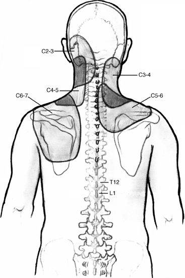
11 I. Clinical categories A. Discogenic axial pain with or without referred pain B. Disc herniation 1. Myelopathy 2. Radiculopathy C. Cervical spondylosis 1. Radiculopathy (foraminal stenosis) 2. Myelopathy II. History and examination A. Cervical radiculopathy 1. Dermatomal pain distribution (Fig. 11–1) a. Spurling’s sign (1) Pain exacerbated by neck extension and rotation toward the symptomatic side b. Shoulder abduction relief sign (1) Pain ameliorated by neck flexion and shoulder raise 2. Neurological findings (nerve root distribution) a. Numbness b. Paresthesias c. Weakness d. Hyporeflexia B. Cervical myelopathy 1. Pain is usually absent. a. Discomfort varies from a dull ache to sharp pain. Figure 11–1 Neck pain and referred pain from the cervical zygapophyseal joints. 2. Symptoms a. Wide, ataxic gait pattern b. Poor hand dexterity (1) Buttoning shirt (2) Writing (3) Holding onto a coffee mug 3. Physical exam findings a. Hyperreflexia b. Positive Hoffman’s sign c. Positive Babinski
Degenerative Cervical Spine Disorders: Surgical and Nonsurgical Treatment
♦ Cervical Degenerative Disease

| Cervical Spondylosis | Disk Herniation | |
| Age | >50 | <50 |
| Sex | Male > female | Male = female |
| Onset | Insidious | Acute |
| Location of pain | Neck and arm | Arm |
| Neck stiffness | Yes | No |
| Weakness | Yes | Yes or no |
| Myelopathy | More common | Less common |
| Dermatomal distribution | Multiple | Single |
d. Positive Lhermitte’s sign
e. Myelopathic hand syndrome
(1) Thenar atrophy
(2) Positive finger escape sign
(3) Positive grip release test
(4) Dysdiadochokinesia
(a) Loss of coordination and dexterity of the hands during rapid movement (Table 11–1)
III. Radiographic imaging (Figs. 11–2, 11–3)
A. Plain radiographs
1. Anteroposterior, lateral, and oblique views
a. Overall alignment
(1) Patients with spondylosis will have a loss of lordosis or spondylolisthesis
b. Narrowing of the intervertebral disc space
c. Degenerative changes in the zygapophyseal joints and the presence of osteophytes
d. Foraminal narrowing observed on the oblique views (Fig. 11–4).
B. Myelography and computed tomography myelography
1. Modality of choice for those who cannot undergo magnetic resonance imaging (MRI)
2. Good for postoperative imaging if instrumentation present
3. Disadvantage is procedure invasiveness
C. MRI
1. Imaging modality of choice for cervical disc disease
2. Good for evaluating space available for the cord
a. Less than 13 mm is relative stenosis.
b. Less than 10 mm is critical stenosis.
3. Particularly useful to rule out spinal cord lesions such as syringomyelia, tumors, and myelomalacia
4. Correlation with clinical symptoms is critical as the false-positive rate is high.


