Chapter 85 Degenerative Rotatory Scoliosis
Three- Dimensional Thoracic and Lumbar Spine Deformity Correction
Degenerative scoliosis is the most common cause of adult scoliosis.1 It develops de novo during adulthood and is largely due to asymmetrical disc degeneration; the resultant curve has even been referred to as a “discogenic curve.”1 Additionally, it may be related to osteoporosis and associated compression fractures. The apex of this curve is most often present at L2-3 or L3-4 and is usually limited to the lumbar or thoracolumbar regions. Its extent, as illustrated by imaging studies, does not necessarily correlate with symptoms or neurologic deficits, a fact that presents a significant dilemma to the treating physician. Management options are complicated by the wide variety of treatment choices.
Pathophysiology of Disc Degeneration and the Spondylotic Process
Lumbar spondylosis is not a pathologic process; it is but a manifestation of the wear and tear associated with aging, specifically the consequences of loading. It is defined as vertebral osteophytosis secondary to degenerative disc disease2 and is not an inflammatory process. Noninfectious inflammatory processes are grouped together as arthritides and are excluded from this discussion.
Spondylosis and associated osteophytosis are universally accompanied by degeneration of the intervertebral disc. The intervertebral disc is an amphiarthrodial joint (no synovial membrane) with particular traits that result in a characteristic degeneration pattern. Conversely, arthritides classically involve the synovial membranes of diarthrodial joints (joints lined with synovium and lubricated with synovial fluid, such as facet joints). Facet joints, however, are also affected by the spondylotic process.3,4
The degenerative process primarily involves the disc interspace and alters intradiscal dynamics that result in spine deformation. The resultant excessive motion and stresses cause extradiscal soft tissue proliferation. Finally, spine deformation predisposes to further deformation (see the section titled “Osteoporosis”). Osteoporosis contributes to the latter process, with a resultant asymmetrical vertebral body collapse.
Intradiscal Dynamics
The water content of the disc interspace gradually decreases throughout life, which contributes to alterations in the chemical and anatomic makeup of the disc. Fibroblasts become defective, and the desiccated disc is less effective as a cushion. Fissures then develop in the cartilaginous end plates. Schmorl nodes are manifestations of this pathologic process. Gas may accumulate in the disc (the vacuum phenomenon). An ingrowth of fibrocartilage (mucoid degeneration) with obliteration of the nucleus fibrosus ensues. Relative incompetence of the disc itself and relative instability result, and anulus fibrosus bulging and tension occur as a result of this process.3
Disc Deformation
Form follows function, even during the process of degeneration. Therefore, osteophyte formation occurs predominantly on the concave side of a scoliotic curvature (where anulus fibrosus bulging is most significant), while disc herniation occurs commonly on the convex side of a spinal bend. The thin dorsal anulus fibrosus and relatively weak lateral aspect of the posterior longitudinal ligament combine with the migratory tendencies of the nucleus pulposus to encourage dorsolateral disc herniation.3
In the laboratory, (1) flexion (causing dorsal nucleus pulposus migration), (2) lateral bending away from the side of disc herniation (causing lateral nucleus pulposus migration), and (3) application of an axial load (causing an increase in intradiscal pressure) are required for the creation of a herniated lumbar disc. A degenerated disc is also necessary as a predisposing factor.5 This complex loading pattern results in the application of tension on the weakest portion of the anulus fibrosus (the dorsolateral position, the location of the herniation), migration of the nucleus pulposus toward this position, and an asymmetrical increase in intradiscal pressure. The age-related increased frequency of anulus fibrosus tears and a peaking of nucleus fibrosus pressures in people 35 to 55 years of age4 also predispose to an increased incidence of disc herniation. Asymmetrical collapse of the disc interspace is often a result of the disc degeneration process and places asymmetrical focal stresses on portions of the spine.
Spinal Configuration
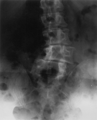
The coupling phenomenon (whereby one movement of the spine about or along an axis obligates another movement about or along another axis)3 plays a significant role in the development of degenerative spine deformations in the lumbar region (whereas it is of minimal significance regarding degenerative deformities in the thoracic region). This is because thoracic degenerative deformities are often oriented in the sagittal plane, whereas degenerative lumbar deformities are usually oriented in the coronal plane (excluding degenerative lumbar spondylolisthesis). The absence of uncovertebral joints (in contrast to the cervical region) and the sagittal orientation of the facet joints (in contrast to the cervical and thoracic regions) create a situation that causes obligatory rotation of the spine in response to lateral bending (coupling) and, commonly, a loss of normal lumbar lordosis. The progression of lateral bending deformities in the lumbar spine (scoliosis) thus predisposes to rotation of the spine (Fig. 85-1), and the influence of an uncompensated thoracic kyphosis predisposes the lumbar spine to greater “flattening” or loss of the normal lordotic curve.
Not all scoliotic curves are symptomatic, as patients may be able to compensate for these deformities by “rebalancing” the spine through other skeletal structures, such as pelvic tilt. When curve progression can no longer be compensated, the subsequent displacement of the load causes worsening of curve that caused it in the first place. Therefore, lateral bending deformation predisposes to lateral bending deformity progression in the lumbar spine, as the presence of kyphotic deformation predisposes to the progression of kyphotic deformation in the thoracic spine. An asymmetrical loss of height of the lumbar intervertebral disc may progress to an asymmetrical collapse of the vertebral body, as described previously in this chapter. As this scoliotic deformity progresses, it is obligatorily associated with rotation of the spine, with the spinous processes rotating toward the concave side of the curve (coupling).3 Of note is that because of the aforementioned osteophyte development propensities, osteophytes occur predominantly on the concave side of the curvature.
Operative Treatment
The operative treatment of scoliosis is reserved for patients with refractory pain due to the scoliosis curve, significant curve progression, gait disturbance, and neurologic deficit all leading to a significant limitation of activities of daily living.6,7 Preoperative preparation should include adequate imaging, as was already mentioned.
Any patient being considered for surgery not should only get detailed radiographic spine imaging but may also need a variety of complementary studies. A dual-energy x-ray absorptiometry scan can provide useful information about bone quality that may affect surgical planning. Patients with suspected pulmonary compromise should be sent for pulmonary function testing, although pulmonary compromise is rare in patients with curves less than 80 degrees.8 Medical and cardiac risk stratification should be obtained for anyone with a significant medical history. It should be noted that occult cardiac disease can be seen in adult scoliotic patients owing to severe deconditioning and the patient’s inability to experience exercise-related stress. Smoking cessation should be pursued, and a general rule of thumb is that elective surgery for deformity correction be offered to patients only after they have quit smoking.
The current approach to the surgical treatment of scoliosis is primarily pedicle screw and rod instrumentation. A recent report comparing hook-rod constructs and pedicle screw-rod constructs found that no pedicle screw patient required revision surgery for instrumentation-related complications and, overall, pedicle screw patients were 89% less likely to require revision surgery.9 These patients were also found to have better curve correction and maintenance of thoracic kyphosis, and pedicle screw-rod constructs often negated the need for ventral release surgery.10 However, hooks remain a valuable alternative when pedicle screws are contraindicated.
Goals of surgical correction of scoliosis are correction of coronal and sagittal balance to decrease pain, to decompress the neurologic elements, to correct balance so as to improve function, and to provide cosmesis.11
Related posts:


Stay updated, free articles. Join our Telegram channel

Full access? Get Clinical Tree


