Design and Surgical Technique of the FlexiCore Lumbar Artificial Disc
Philippe Lauweryns
Design
The FlexiCore Intervertebral Disc is comprised of four components, which are assembled by the manufacturer and provided to the surgeon as a single unit (Fig. 22.1). The four components are as follows:
A highly polished semi-spherical head
A superior baseplate having a domed upper surface and a cylindrical stem extending from the lower surface and into the head
An inferior baseplate having a domed lower surface and a semispherical recess formed in the upper surface to accommodate the head
A shield seated around the head and press fit to the upper surface of the inferior baseplate
The exterior surfaces of the superior and inferior baseplates each have a central dome that rises 2 mm at its peak to ensure proper seating of the device between the endplates of the adjacent vertebral bodies. Two sets of 1.5-mm spikes are provided on each baseplate for additional bone fixation.
The principal articulating components of the FlexiCore Intervertebral Disc are the semispherical head and the semispherical recess in the upper surface of the inferior baseplate. These components are located directly between the domes on the exterior surfaces of the baseplates, providing a center of rotation similar to that of a healthy natural disc.
The FlexiCore Intervertebral Disc was designed to allow for repositioning in situ. Three holes pass through the edge of the shield and into the upper surface of the inferior baseplate and enable anterior, anterolateral right, or anterolateral left attachment of the inserter/impactor and repositioner/extractor instruments. Four holes in the superior baseplate can be engaged for implant repositioning or removal.
The implant is comprised of cobalt chromium molybdenum alloy (ASTM F-1537). To promote fusion with the vertebral body endplates, the external surfaces of the superior and inferior baseplates are plasma sprayed with a layer of CP titanium.
The FlexiCore Intervertebral Disc is available in two baseplate areas (28 ÷ 35 mm and 30 ÷ 40 mm) and seven heights (12, 13, 14,
15, 16, 17, and 18 mm). The overall height of each implant is measured between the base of the spikes on the superior and inferior baseplates when the device is in the parallel (nonlordosed) position.
15, 16, 17, and 18 mm). The overall height of each implant is measured between the base of the spikes on the superior and inferior baseplates when the device is in the parallel (nonlordosed) position.
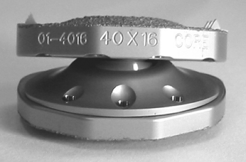 FIGURE 22.1 FlexiCore Intervertebral Disc. |
The range of motion of the implant is designed to be 32 degrees (at least 30 degrees) in both flexion/extension and lateral bending when the device is in the neutral position. The neutral position is nonlordosed and at the midpoint of those articulations. Axial rotation between the baseplates is designed to be 14 degrees (at least 10 degrees) when the device is in the neutral position. Range of motion in situ is limited by soft tissue balance and the mechanical stops provided by the facet joints of the individual subject.
The FlexiCore Intervertebral Disc incorporates a semispherical articulating surface that maintains a fixed rotation point centered between the peaks of the domes of the baseplates. Because the domes fit within the concavities of the vertebral body endplates, the device’s center of rotation is biased toward a location that causes the modes of motion of the spinal segment to be similar to those of a healthy natural segment.
The FlexiCore Intervertebral Disc is indicated for the treatment of lumbar discogenic pain unresponsive to conservative treatment associated with degenerative disc disease (DDD). The FlexiCore Intervertebral Disc is intended to replace a degenerated lumbar intervertebral disc (L1-S1), permit motion of the treated segment, improve function, and reduce back pain associated with DDD.
The primary goal of the disc arthroplasty consists of, but is not limited to, restoring or preserving the segmental motion of the intervertebral joint(s) (1,3,11,16,19,22,38). The total disc replacement (TDR) is aimed as well at reducing stresses on the adjacent level in the hope of reducing adjacent level degeneration (1,11,16,19,38). The facet joints must be protected from further degeneration (22). Segmental stability, disc height and sagittal alignment are expected to be provided as well (16,22). The overall goal is to relieve pain and to get the patient back to normal activity and work (1,16,38).
Ideally some designers looked at reproducing the native disc performances. But basically two different designs came out: some designs aimed to reproduce the viscoelastic properties of the disc, whereas others aimed at the reproduction of “only” the motion characteristics of the disc. The latter design was mostly inspired from the peripheral joints prostheses (10,38). Due to the sensitive location of a disc prosthesis it is commonly agreed the device must be safe so that no catastrophic consequence and vessels injury may happen when it fails (17). It is expected that the disc replacement device must protect the posterior elements, that is the facet joints for long-term results (12,22). There is one more important feature of the design: Because the anterior stabilizing structures are disrupted during the implantation, the device must be self stable, with primary and secondary fixation characteristics (22).
The design of an artificial disc may include some constraints in the design to limit the range of motion. No standard has been made yet but one can distinguish three concepts: unconstrained, semiconstrained, or a fully constrained design (1).
It is expected an unconstrained device would more likely provide a physiologic center of rotation (22,35), to be less sensitive to error of placement (22), and to have the longest survival (32), but at the risk of transferring more rotational load on the facet joints (35). Constrained designs include a fixed center of rotation by means of a fixed polyethylene (PE) core (Prodisc) or a ball-in-socket design (Maverick-Flexicore). Constraints may aim in addition at protecting the posterior elements from increased loads (22,26).
As the native disc has a function of creeping and relaxation (23) various materials have been suggested. With the exception of the rubber material included in some discs, it has been demonstrated that the most common materials do have the same very
limited shock absorption capacity. It has been generally believed that softer material like PE will provide some cushioning, but tests showed that PE-based devices and metal on metal (MoM) devices both have a similar behavior and no significant shock absorption capacity (29).
limited shock absorption capacity. It has been generally believed that softer material like PE will provide some cushioning, but tests showed that PE-based devices and metal on metal (MoM) devices both have a similar behavior and no significant shock absorption capacity (29).
Considering the age of patients suffering from chronic low back pain it is usually expected a TDR to be viable for 40 years (17,26,40). The SB Charité prosthesis has been tested for 20 million cycles only at 8000 N without cracks (4), but cracks and wear have been reported in clinical use (39). Kostuik et al. (26) suggested a test for an implantable device to be up to 100 million cycles from which 85 million standard cycles would represent the claimed 40 years of life (17,26). To assess its durability, the MoM Maverick device has been successfully tested in a different model simulating more than 31 years of activity (30).
Due to the extended exposure and the extensive annulotomy necessary to insert a disc, the primary and secondary fixation are critical issues to consider. Various designs/techniques have been used with success so far, but the available follow-up is of a maximum of 17 years (36). The surgical technique differs from a standard anterior lumbar interbody fusion (ALIF): Stability cannot be provided by excessive disc distraction, which would limit the motion and painfully stretch the nerve roots (14). Some TDR designs rely on a keel inserted into the adjacent vertebral body to be stable in the short term, that is, Prodisc and Maverick (14,30). Other designs rely on small teeth on the device endplate to anchor the TDR device into the soft bone (SB Charité) (4). Both concepts showed in clinical use a fairly reliable fixation in the immediate postoperative period. Keels look intuitively very effective and safe but they mandate skilled hands and a very accurate preparation before insertion, because no per operative adjustment is possible (14).
Long-term fixation is biologic and calls for a tight bone bonding onto the device endplate. Various surface coatings are used: titanium, hydroxy apatite, and plasma spray (7,14,30,37,41) of which the effectiveness has been demonstrated in the hip and knee prosthesis experience.
Related posts:
 Adjacent Level Disc Biomechanics
 Nucleus Pulposus Regeneration: Present Limitations and Future Opportunities
 Prosthetic Disc Nucleus: Treatment with the Anterior Approach
 Total Lumbar Disc Arthroplasty: Overview of Clinical Results for Existing Implants
 Motion Preservation—Disc Replacement for Lumbar Degenerative Disorders with the ProDisc®-L Prosthesis
 Posterior Nonfusion Stabilization of the Degenerated Lumbar Spine with Cosmic
Stay updated, free articles. Join our Telegram channel

Full access? Get Clinical Tree


