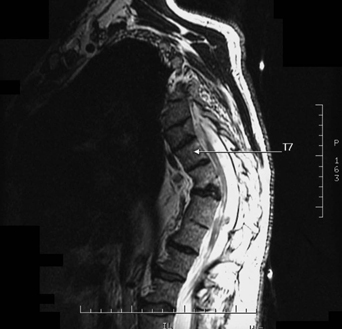
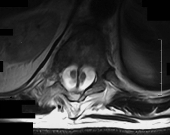
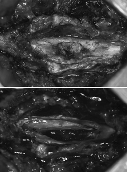
79 A 59-year-old man presented with progressive kyphoscoliotic deformity of the thoracolumbar spine. He was neurologically intact. Further investigation found diastematomyelia. The patient’s T2-weighted sagittal and axial thoracic magnetic resonance imaging (MRI) had a split cord malformation (Figs. 79-1 and 79-2). FIGURE 79-1 Sagittal thoracic MRI demonstrates diastematomyelia. Split cord malformation type II He had a thoracic laminectomy with removal of an intradural spur and cord untethering and segmental fusion, as demonstrated in pre- and post-spur removal intraoperative pictures (Fig. 79-3). Diastematomyelia is a spinal dysraphic state resulting in congenital splitting of a segment of the spinal canal. FIGURE 79-2 Axial MRI demonstrates diastematomyelia, with a bony ridge entering a single dural sac.
Diastematomyelia
Presentation
Radiologic Findings
Diagnosis
Treatment
Discussion
Diastematomyelia
Only gold members can continue reading. Log In or Register to continue


Full access? Get Clinical Tree


