Fig. 23.1
SIH. Axial T2-weighted image and coronal T1-weighted image after contrast medium administration (a) and (b) demonstrated bilateral subdural fluid collections (arrows) and diffuse dural thickening and enhancement along both cerebral convexities, the falx and the tentorium. Sagittal T1-weighted image with contrast medium (c) shows sagging brain with midbrain caudal displacement and pituitary enlargement. Axial T1-weighted image with contrast medium at the level of L5 (d) demonstrates collapse of the dural sac with engorgement of the epidural venous plexus, and myelo-CT (e) shows CSF leaks (arrowheads)
Spinal studies are performed to determine the exact location of the CSF leak.
Spinal MRI demonstrates three characteristic findings: (1) epidural fluid collections, (2) collapse of the dural sac with engorgement of the epidural venous plexus, and (3) abnormalities of the root sleeves (Fig. 23.2). The epidural collections correspond to accumulations of CSF leaking into the epidural space [7]. Myelo-MR, radioisotope cisternography with Indium-111, and myelo-CT may reveal meningeal diverticula or Tarlov’s cysts that often represent the site of the leak (Fig. 23.1).


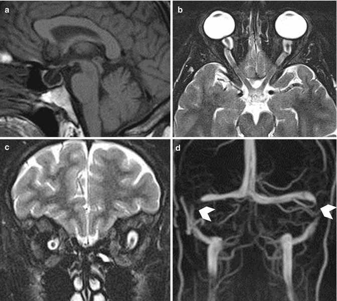
Fig. 23.2
IIH. Sagittal T1-weighted image (a) shows a huge empty sella. Nerve tortuosity, nerve sheath distention, and flattenting of the globe and papilla in globe are demonstrated in axial and coronal T2-weighted images (b, c). In venous MR angiography stenosis, in the middle portion of bilateral transverse sinuses (arrowheads) is visible (d)
23.1.6 Differential Diagnosis
The herniation of the cerebellar tonsils in SIH may simulate Chiari I malformation.
Meningeal dural enhancement may be confused with meningeal carcinomatosis.
At MRI, hypertrophic pachymeningitis may be indistinguishable from SIH.
Enlargement of the pituitary gland due to hyperemia may mimic pituitary tumors.
23.1.7 Treatment and Prognosis
Available treatment options are largely empiric; none have been evaluated in randomized, placebo-controlled clinical studies [4].
Initial therapy consists of strict bed rest supported by adequate oral hydration.
Spontaneous remissions within 2–6 months are observed in about 40 % of patients [8].
If further treatment is required, autologous lumbar epidural blood patch (EPB) is the first choice. With EPB, 61–75 % of patients (more than 90 % in the author’s personal experience) improve.
Improvement may be obtained even months after the onset of pain [9].
Of 25 consecutive patients with spontaneous CSF leaks treated with EBP, nine patients (36 %) responded well to the first injection. Of 15 patients who received a second EBP, five became asymptomatic (33 %). Of eight patients who received three or more EBP (mean 4), four patients (50 %) showed symptoms remission. Bilateral chronic subdural hematoma caused by CSF leak had to be evacuated in two patients (5 %) [8].
In a large unpublished series (Enrico Ferrante, personal communication. E-mail: enricoferrante@libero.it), regarding 210 SIH patients (111 women and 99 men) submitted to blood patch and followed-up from 6 months to 8 years, 198 had a positive response, 10 patients were treated two or more times and only two did not achieve symptomatic remission.
Similar results were obtained in a group of 196 patients with SIH; 130 had a spontaneous recovery within 6 months. In 66 of the remaining cases, 58 recovered after one or more blood patches. In 4 cases, recovery was obtained only after surgery; in 4 patients SIH became chronic (author’s personal experience).
The response to a single EBP may not be permanent, and complete relief of symptoms may only occur after two or more procedures.
Fibrin sealant may be considered as a second choice option.
Surgical intervention at the site of leak – if found – is safe and commonly successful in eliminating the cause of the leakage. In a minority of patients (1–2 %), all therapeutic attempts fail; therefore, patients may suffer chronic symptoms and work disability.
Dementia and cognitive deficits are uncommon complications [1].
Although unusual, parkinsonism, ataxia, and/or stupor and coma have also been described.
23.2 Idiopathic Intracranial Hypertension (IIH)
Key Facts
Terminology – IIH is a syndrome of increased intracranial pressure without identifiable cause and with normal CSF
Clinical features
–Diffuse headache aggravated by physical activity and associated with nausea and vomiting.
–Papilledema
Diagnostic markers
CSF
–Elevated opening pressure reaching more than 200 mmH2O in nonobese and more than 250 mmH2O in obese patients
Imaging – MRI is the study of choice, showing
–Optic nerve distension
–Empty sella with deformities of the pituitary gland; distension of the optic nerve sheath
–Posterior globe flattening and tortuosity of the optic nerve
–MRV shows venous narrowing or cerebral venous sinus thrombosis
Pathogenesis
Causes are largely unknown
Obesity, delayed CSF absorption, and venous outflow abnormality are involved in the elevation of intracranial pressure
Stenotic transverse sinuses (TSS) have been observed in up to 90 % of IIH patients
Top differential diagnoses
Migraine and tension-type headache
Optic disc swelling caused by different etiologies
Obstructive hydrocephalus
Prognosis
IIH is a chronic disease that requires long-term treatment.
Permanent, mild, or moderate visual field constriction has been reported in the follow-up.
Prognosis is good in patients who are promptly diagnosed and receive appropriate treatment.
Recurrence is rare.
23.2.1 Terminology and Definitions
Idiopathic intracranial hypertension (IIH) (alias: pseudotumor cerebri, benign intracranial hypertension) is a syndrome of increased intracranial pressure with no identifiable cause and with normal CSF composition related to an altered cerebrospinal fluid (CSF) hydrodynamics.
23.2.2 Demographics
The annual incidence is 0.9 cases per 100,000 with a prevalence of 8.6 cases per 100,000. The disorder usually affects obese women of childbearing age. In adults, the female to male ratio ranges between 4.3 and 5.0:1.0 [2].
23.2.3 Clinical Features
The typical IIH patient is an overweight woman of short stature. Diffuse headache, aggravated by physical activity, often associated to nausea and vomiting, is the most common complain. Papilledema, typically present in IIH, may cause scotomas and from transient dimming of vision to complete visual loss. A protracted course, lasting months to years, appears to be common; however, a subset of patients may show a more fulminant course with rapid development of vision loss within a few weeks from onset.
Definite diagnostic criteria for definite pseudotumor cerebri (PTCS) require: (1) papilledema, (2) normal neurologic examination except for cranial nerve abnormalities (usually unilateral or bilateral abducens nerve palsy), (3) normal brain parenchyma on MRI, (4) normal CSF composition, and (5) elevated lumbar puncture opening pressure (250 mm).
In the absence of papilledema, the diagnosis of PTCS can be made if the second and the fifth criteria described above are satisfied. In the absence of papilledema or sixth nerve palsy, a diagnosis of PTCS can be suggested, but not definitively made, if the second and the fifth above criteria are satisfied together with three of the following neuroradiological findings: (1) empty sella, (2) flattening of the posterior aspect of the globe, and (3) distension of the perioptic subarachnoid space with or without tortuous optic nerve, transverse sinus stenosis. A diagnosis of PTCS is definite if the patient fulfils 1–5 criteria. The diagnosis is considered probable if 1–4 criteria are satisfied and the CSF open pressure is lower than specified [10].
23.2.4 Diagnostic Markers
CSF
Elevated CSF opening pressure (more than 200 mmH2O in nonobese patients; more than 250 mmH2O in the obese) is normally found. Increases of intracranial pressure may occur intermittently. Their demonstration may require continuous monitoring in some patients.
MRI
is the study of choice. Signs of elevated intracranial pressure: optic nerve tortuosity and distension, enlargement of its sheath, empty sella, and posterior eye globe flattening are frequently found. Magnetic resonance venography (MRV) may show venous narrowing or venous sinus thrombosis. Venous sinus occlusion and arterio-venous fistulas may produce PTCS [10, 11] (Fig. 23.2).
23.2.5 Pathogenesis
The underlying cause of IIH is largely unknown.
Obesity is present in more than 70 % of adult IIH patients. Body mass index correlates with CSF opening pressure so that weight reduction results in decreased intracranial pressure.
Stenotic transverse sinuses (TSS) have been observed in up to 90 % IIH patients, but there are controversies as to whether TSS cause or are the consequence of raised intracranial pressure.
Reduction of intracranial pressure through CSF subtraction (by lumbar puncture or diversion procedures) may improve IIH.
23.2.6 Differential Diagnosis
Papilledema may have a similar appearance to optic disc swelling of different causes.
Adhesions of arachnoid granulations from infection or subarachnoid hemorrhage may modify CSF reabsorption and cause elevated intracranial pressure (ICP).
Obstructive hydrocephalus should be considered in the differential diagnosis [12].
23.2.7 Treatment and Prognosis
IIH is a substantially chronic disease in which recurrences, sometimes correlated with recent weight gain, are unpredictable.
Stay updated, free articles. Join our Telegram channel

Full access? Get Clinical Tree


