Lesions of the cerebral centers and connections that subserve language function may cause aphasia, an abnormality of language, even though the articulation mechanisms may be intact. Mutism is a total inability to speak; usually the patient appears to make no attempt to speak or make sounds. Mutism is usually of psychogenic origin if present in an apparently otherwise normal patient, but it may occur with lesions of the cerebrum, brainstem, and cerebellum (especially in children). In akinetic mutism, the patient is mute and unmoving (akinetic). The patient appears awake but is mute, immobile, and unresponsive. Akinetic mutism most often occurs with damage to the frontal lobes. Selective (elective) mutism is a disorder of childhood characterized by a total lack of speech limited to certain situations—such as school—despite normal speech in other settings. The present discussion is limited to disorders limited to the motor components of speech; disorders of language are discussed subsequently.
Abnormalities of articulation may be caused by many different pathologic conditions. Disturbances in the respiratory rhythm interfere with speech, and respiratory muscle weakness causes a feeble voice with abnormalities in regularity and rhythm. Laryngeal disease may cause severe speech impairment, but whispered speech may still be possible. In children, articulation disturbances may be developmental and are often temporary. Structural abnormalities of the vocal tract, such as congenital craniofacial defects (cleft palate, cleft lip), ankyloglossia (abnormal shortness of the frenulum of the tongue; “tongue-tie”), adenoidal hypertrophy, vocal cord edema or nodules, nasal obstruction, or perforated nasal septum may cause abnormalities in sound production. The importance of the teeth in articulation is apparent in the speech of edentulous patients.
Neurologic disturbances of articulation may be caused by the following: primary muscle diseases affecting the tongue, larynx, and pharynx; neuromuscular junction disorders; lower motor neuron disease involving either the CN nuclei or the peripheral nerves that supply the muscles of articulation; cerebellar dysfunction; basal ganglia disease; or disturbances of the upper motor neuron control of vocalization. A commonly used classification separates dysarthria into flaccid, spastic, ataxic, hypokinetic, hyperkinetic, and mixed types.
Lesions of the hypoglossal nerve or nucleus—or local disorders of the tongue such as ankyloglossia—may cause impairment of all enunciation, but with special difficulty in pronouncing lingual sounds. The speech is lisping in character and is clumsy and indistinct. Paralysis of the laryngeal musculature causes hoarseness, and the patient may not be able to speak above a whisper; there is particular difficulty pronouncing vowels. Similar changes occur in laryngitis and in tumors of the larynx. With unilateral laryngeal muscle weakness, such as in recurrent laryngeal nerve lesions, the voice is usually low-pitched and hoarse. However, occasionally severe unilateral vocal cord weakness may be present without much effect on speech because the normal vocal cord is able to adduct across the midline and approximate the abnormal cord. Hoarseness due to slight vocal cord weakness may be brought out by having the patient talk with his head turned to one side. With paralysis of the cricothyroid, the voice is hoarse and deep and fatigues quickly. Diplophonia is one sound being produced at two different frequencies because of differences in vibration when one vocal cord is weak and the other normal. In bilateral abductor paresis, speech is moderately affected, but in bilateral total paralysis it is lost.
Paralysis limited to the pharynx causes little detectable impairment of articulation. Weakness of the soft palate results in nasal speech (rhinolalia, Gr. lalia “speech”), caused by inability to seal off the nasal from the oral cavity. Voice sounds have an added abnormal resonance. There is special difficulty with the velar sounds, but labials and linguals are also affected because much of the air necessary for their production escapes through the nose. The speech resembles that of a patient with a cleft palate. Characteristically, b becomes m, d becomes n, and k becomes ng. Amyotrophic lateral sclerosis and MG are common causes of this type of speech difficulty.
Seventh nerve paralysis causes difficulty in pronouncing labials and labiodentals. Dysarthria is noticeable only in peripheral facial palsy; the facial weakness in the central type of facial palsy is usually too mild to interfere with articulation. Bell’s palsy occasionally causes marked dysarthria because of inability to close the mouth, purse the lips, and distend the cheeks. Similar articulatory defects are found in myopathies involving the labial muscles (e.g., facioscapulohumeral or oculopharyngeal dystrophy), in cleft lip and with wounds of the lips. There is little impairment of articulation in trigeminal nerve lesions unless the involvement is bilateral; in such cases, there are usually other characteristics of bulbar speech. Trismus may affect speech because the patient is unable to open the mouth normally.
Lower motor neuron disorders causing difficulty in articulation may occur in cranial neuropathies. Lesions of the ninth and eleventh nerves usually do not affect articulation. A unilateral lesion of CN X causes hypernasality. Lesions involving the vagus bilaterally distal to the origin of the superior laryngeal nerve may leave the vocal cords paralyzed in adduction, resulting in a weak voice with stridor. With more proximal lesions, there is no stridor but the voice and cough are weak.
Neuromuscular disorders, particularly neuromuscular junction disorders, often interfere with speech. In MG, prolonged speaking, such as counting, may cause progressive weakness of the voice with a decrease in volume and at times the development of a bulbar or nasal quality, which may even proceed to anarthria. As the voice fatigues, the speech of a patient with bulbar myasthenia may be reduced to an incoherent whisper. Thomas Willis, who provided one of the first descriptions of MG in 1672, wrote of a woman who, when she tried to talk for a prolonged period, “temporarily lost her power of speech and became mute as a fish.” An occasional myasthenic patient must hold his jaw closed with his hand in order to enunciate.
Motor neuron disease commonly causes dysarthria. The type varies from a primarily flaccid dysarthria in bulbar palsy to a primarily spastic dysarthria in primary lateral sclerosis; most patients have classical amyotrophic lateral sclerosis, and the dysarthria is of mixed type with both flaccid and spastic components; that is, there are both bulbar palsy and pseudobulbar palsy (see below). In bulbar palsy, dysarthria results from weakness of the tongue, pharynx, larynx, soft palate, and, to a lesser extent, the facial muscles, lips, and muscles of mastication. Both articulation and phonation may be affected; speech is slow and hesitant with failure of correct enunciation, and all sounds and syllables may be indistinct. The patient talks as though his mouth were full of mashed potatoes. Speech is thick and slurred, often with a nasal quality and a halting, drawling, monotonous character. The tongue lies in the mouth, more or less immobile, shriveled and fasciculating; the palate rises very little. The dysarthria may progress to a stage where there is phonation but no articulation. Speech is reduced to unmodified, unintelligible laryngeal noises. Often at this stage, the jaw hangs open and the patient drools. The condition may eventually reach the stage of anarthria. Dysphagia is typically present as well. For an audio of flaccid dysarthria see http://www.youtube.com/watch?v=dy8WvykiLto&feature=related.
Supranuclear lesions involving the corticobulbar pathways may also cause dysarthria. Unilateral cortical lesions do not usually affect speech unless they are in the dominant hemisphere and cause aphasia. Occasionally some dysarthria accompanies aphasia. Rarely, lesions in the cortical motor areas for articulation may cause severe dysarthria without aphasia. Both dysarthria and dysprosody, a defect in rhythm, melody, and pitch, have been described with localized frontal lobe lesions; these may be due to an apraxia of speech (AOS). In acute hemiplegia, there may be transient slurring or thickness of speech depending on the degree of face and tongue weakness.
Bilateral supranuclear lesions involving the cortex, corona radiata, internal capsule, cerebral peduncles, pons, or upper medulla may cause pseudobulbar palsy with spastic dysarthria. The muscles that govern articulation are both weak and spastic. Phonation is typically strained-strangled, and articulation and diadochokinesis are slow. There is a thick bulbar type of speech, similar to that in progressive bulbar palsy, but more explosive; it rarely progresses to complete anarthria. The tongue is protruded and moved from side to side with difficulty. There may also be spasticity of the muscles of mastication; mouth opening is restricted and speech seems to come from the back of the mouth. The jaw jerk, gag reflex, and facial reflexes often become exaggerated and emotional incontinence commonly occurs (pseudobulbar affect). For a video of spastic dysarthria see http://www.youtube.com/watch?v=EHNSBo3SsmY. The Foix-Chavany-Marie (bilateral anterior opercular) syndrome is the loss of voluntary bulbar movements, with preservation of involuntary movements and reflexes, due to a lesion involving the frontal opercular regions bilaterally. Unilateral lesions of the dominant frontal operculum may cause “cortical dysarthria” or AOS (see below).
Lesions of the basal ganglia may affect speech. Athetotic grimaces of the face and tongue may interfere with speech. Irregular spasmodic contractions of the diaphragm and other respiratory muscles, together with spasms of the tongue and pharynx, may give the speech a curious jerky and groaning character. In addition, there may be a pseudobulbar element with slurred, indistinct, spastic speech. When chorea is present, the violent movements of the face, tongue, and respiratory muscles may make the speech jerky, irregular, and hesitant. The patient may be unable to maintain phonation, and occasionally there is loss of the ability to speak. Dysarthria is one of the most common neurologic manifestations of Wilson’s disease, and frequently the presenting complaint. It is typically mixed with spastic, ataxic, hypokinetic, and dystonic elements. The type of dysarthria often corresponds with other manifestations, with spasmodic dysphonia in those with dystonic features, hypokinetic in those with parkinsonism, and ataxic in those with tremor as the primary manifestation. Pantothenate kinase-associated neurodegeneration (Hallervorden-Spatz syndrome) may cause a similar mixed spastic-extrapyramidal dysarthria.
Speech in parkinsonism is often mumbled, hesitant, rapid, and soft (hypophonic). Parkinsonian patients tend to be soft, fast, mumbly talkers. There may sometimes be bradylalia, with feeble, slow, slurred speech because of muscular rigidity and immobility of the lips and tongue. There is dysprosody and the speech lacks inflections, accents, and modulation. The patient speaks in a monotone, and the words are slurred and run into one another. The voice becomes increasingly weak as the patient talks, and he may become unable to speak above a whisper; as the speech becomes more indistinct it may become inaudible or practically disappear. Words may be chopped off. There may be sudden blocks and hesitations, or speech may stop abruptly. There may be pathologic repetition of syllables, words, or phrases (palilalia). Like the parkinsonian gait, the speech may show festination, with a tendency to hurry toward the end of sentences or long words.
Voice tremor produces rhythmic alterations in loudness and pitch. There may be associated tremor of the extremities or head, or other signs of neurologic dysfunction. Voice tremor may further complicate the other speech disturbances of parkinsonism. Voice tremor occurs commonly in essential tremor, a frequently familial syndrome that most often affects the hands. Fine voice tremors are characteristic of essential tremor; coarse tremors are more commensurate with cerebellar disease. Essential voice tremor is probably more common than generally suspected, and many cases appear to go unrecognized or misdiagnosed, most often as spasmodic dysphonia. Voice tremor is a common manifestation of anxiety. Lip and chin tremors, when severe, may interfere with speech.
In habit spasms, Tourette’s syndrome, and obsessive-compulsive states, there may be articulatory tics causing grunts, groans, or barking sounds. In Tourette’s syndrome, palilalia may also occur.
Cerebellar dysfunction causes a defect of articulatory coordination (scanning speech, ataxic dysarthria, or speech asynergy). Many studies have attempted to localize speech functions in the cerebellum. The superior regions bilaterally appear to mediate speech motor control and the right cerebellar hemisphere has a putative role in speech planning and processing. Lesion mapping studies have shown that dysarthria occurs with pathology affecting the upper paravermal areas, or lobules V and VI. Subtypes of ataxic dysarthria are recognized, common to all is an impairment of articulation and prosody.
Ataxic dysarthria causes a lack of smooth coordination of the tongue, lips, pharynx, and diaphragm. Ataxic speech is slow, slurred, irregular, labored, and jerky. Words are pronounced with irregular force and speed, with involuntary variations in loudness and pitch lending an explosive quality. There are unintentional pauses, which cause words and syllables to be erratically broken. Excessive separation of syllables and skipped sounds in words produce a disconnected, disjointed, faltering, staccato articulation (scanning speech). The speech pattern is reminiscent of a person who is sobbing or breathing hard from exertion. The unusual spacing of sounds with perceptible pauses between words and irregular accenting of syllables may cause a jerky, singsong cadence that resembles the reading of poetry. Ataxic speech is particularly characteristic of multiple sclerosis. It may be accompanied by grimaces and irregular respirations. Ataxia of the voice and scanning speech may be more apparent when the patient repeats a fairly long sentence.
Specific speech abnormalities may occur in various neurologic conditions. The disturbance varies in individual cases and depends upon the site of the predominant pathologic change. In multiple sclerosis, the speech is characteristically ataxic; there are explosive and staccato elements, with slowness, stumbling, halting, slurring, and a cerebellar type of speech ataxia. Spastic-ataxic and mixed dysarthrias are also common. In Friedreich’s ataxia, the ataxic, staccato, and explosive elements predominate. Speech is clumsy, often scanning, and the pitch may suddenly change in the middle of a sentence. In alcohol intoxication, the speech is slurred and indistinct. There is difficulty with labials and linguals, and there may be tremulousness of the voice. Conversation is often characterized by a tendency to garrulousness. The patient may repeatedly use words he can pronounce correctly, avoiding the use of other words. This results from loss of cerebral cortical control over thought and word formulation and speech, rather than from a primary articulatory disturbance. In delirium tremens, the speech is tremulous and slurred. Other types of intoxication also produce speech that is thick and slurred. Rarely, the inability to relax muscles in myotonia causes slight speech impairment. Myxedema may cause a low-pitched, harsh, husky, slow, and monotonous voice. General paresis may cause a tremulous, slurring type of dysarthria, with special difficulties with the linguals and labials. Letters, syllables, and phrases are omitted or run together. The speech is slovenly, with ataxia, stumbling, and alliteration, often accompanied by tremors of the lips, tongue, and face.
Patients with some forms of aphasia, dysarthria, dysprosodia, and speech apraxia may begin to sound as if they have developed an unusual accent. The foreign accent syndrome during recovering facial diplegia made one patient from Virginia sound for several months as if she were a Bavarian countess. The foreign accent syndrome has been reported as the only manifestation of a cortical lesion, and as the presenting manifestation of primary progressive aphasia (PPA).
Spasmodic dysphonia is a focal dystonia characterized by a striking abnormality of voice production. In adductor dysphonia, irregular involuntary spasms of the vocal muscles cause erratic adduction of the cords. As the patient strains to speak through the narrowed vocal tract, his voice takes on a high-pitched, choked quality that varies markedly during the course of a sentence. It is most marked in stressed vowels. The dysphonia may lessen or disappear when the patient sighs or whispers. For an audio of spasmodic dysphonia see http://www.youtube.com/watch?v=-cSMezYQx0E&feature=related. The much rarer abductor spasmodic dysphonia causes excessive abduction of the involved cord, and the voice is hoarse and breathy. In both types, there is often a dramatic improvement in the voice during shouting, whispering, or singing. The difference in adductor and abductor spasmodic dysphonia is nicely demonstrated in the audio/video posted by Reich and Meyer. Both types of may respond dramatically to the injection of botulinum toxin into the involved muscle.
Dyslalia may be caused by damage to or structural abnormalities of the vocal tract, such as wounds of the lips, tongue, palate, or floor of the mouth; maxillofacial injuries; perforation of the palate; congenital cleft lip and cleft palate; enlarged tonsils and adenoids; ankyloglossia; and dental malalignment.
Secondary speech disturbances may also occur without abnormalities or specific dysfunction of the articulatory apparatus, as seen in individuals with hearing defects, delayed physical development, mental retardation, and psychogenic disturbances. Severe hearing loss, especially when it occurs early in life before speech patterns are ingrained, can result in abnormalities of speech. The nature and severity of the speech abnormality depend largely upon the degree of hearing loss, the time at which it occurred, and the individual’s ability to compensate. The speech disorder may range from a mild abnormality of articulation to the indistinct and often unintelligible speech of deaf-mutism. A child with slow physical development or psychological problems may retain childish speech until later years. Childish speech may persist in mild mental retardation. In moderate retardation, speech develops late, and the vocabulary is limited. It may be slow, labored, indistinct, and difficult to understand. In the severely retarded, speech is babbling and grunting in character, with a tendency toward echolalia. In delayed puberty and in eunuchism, the male voice retains juvenile or feminine characteristics, while in the virilized woman it may be low-pitched and coarse.
Stuttering (spasmophemia) refers to faulty, spasmodic, interrupted speech characterized by involuntary hesitations in which the speaker is unable to produce the next expected sound. The flow of speech is broken by pauses during which articulation is entirely arrested. Stammering may happen to anyone in certain circumstances, as with embarrassment. Stuttering implies a more severe disturbance of speech, with faltering or interrupted speech characterized by difficulty in enunciating syllables and joining them together. Interference with communication may be profound and the social consequences severe. Stuttering speech is stumbling and hesitant in character, with habitual and spasmodic repetitions of consonants or syllables, alternating with pauses. There may be localized cramps, spasms, and tic-like contractions of the muscles essential to articulation, which may be accompanied by grimaces, spasms and contractions of the muscles of the head and extremities, and spasm and incoordination of the respiratory muscles. The individual may be unable to pronounce certain consonants, with particular difficulty in using dentals and labials. Often the first syllable or consonant of a word is repeated many times. The individual may remain with his mouth open until the articulatory spasm relaxes, then the words explode out until the breath is gone. He then takes another breath, and the process is repeated. Stuttering is markedly influenced by emotional excitement and by the presence of strangers. In spite of difficulty in speaking, the individual may be able to sing without hesitation. There have been accomplished professional singers who stuttered severely in ordinary speech. Britain’s King George VI stuttered severely, as memorably depicted in the motion picture The King’s Speech. Many theories have been offered regarding the etiology of stuttering.
In lalling (lallation, “baby talk”), the speech is childish, babbling, and characterized by a lack of precision in pronouncing certain consonants, especially the letters r and l. A uvular is substituted for a lingual-palatal r, so that “broken reed” is pronounced “bwoken weed.” The diphthong ow or other sounds may be substituted for the l sound, or sometimes l may be substituted for r. T and d may be substituted for s, g, and the k sound. Lalling may occur because of hearing defects, mental or physical retardation, or from psychogenic disorders. In lisping, the sibilants are imperfectly pronounced, and th is substituted for s; a similar defect in articulation may be associated with partial edentulism. Lalling and lisping are usually due to imperfect action of the articulatory apparatus (as in children), persistent faulty habits of articulation, imitation of faulty patterns of articulation, poor speech training, habit, or affectation.
NONORGANIC SPEECH DISORDERS
Emotional and psychogenic factors influence articulation. Speech, but not language, disorders may occur on a nonorganic basis. Nonorganic voice disorders can take many different forms and can be caused by a variety of factors. The most common nonorganic voice disorders are dysphonia and aphonia. Dysarthria, lalling, stuttering, mutism, or anarthria occurs rarely. There may be infantile language wherein the objective pronoun is used as the subject (e.g., “Me want to go home”). Onset is often abrupt, perhaps in association with emotional trauma; there may be periods of remission, and the condition may suddenly disappear. The speech defect may vary in type from time to time. It is often bizarre, and does not correspond to any organic pattern. The patient may fail to articulate and speak only by whispering. Speech may be lost but the patient is able to sing, whistle, and cough. There may be associated dysphagia and globus hystericus.
In anxiety and agitation the speech may be broken, tremulous, high-pitched, uneven, and breathless. Stuttering and stammering are common. The speech may be rapid and jumbled (tachyphemia or tachylalia), or there may be lalling or mutism. In hysterical aphonia, there is profound speech difficulty but no disturbance of coughing or respiration. Manic patients may have a rapid flow of words (pressured speech), often with an abrupt change of subject. In depression speech may be slow, sometimes with mutism. True organic aphasia is occasionally confused with hysterical or simulated mutism. The aphasic patient, no matter how speechless, at least occasionally tries to speak; in hysterical mutism there may be the appearance of great effort without the production of so much as a tone; in simulated mutism the patient does not even make an effort. Mutism may also occur in catatonia. In schizophrenia there may be hesitancy with blocking, or negativism with resulting mutism (alalia). Two common nonorganic dysphonias seen in children and adolescents are the whispering syndrome, seen primarily in girls, and mutational falsetto (hysterical high-pitched voice), seen primarily in boys.
Palilalia, echolalia, and perseveration are often manifestations of psychosis, but they can occur with organic lesions, especially of the frontal lobes. Palilalia is the repetition of one’s own speech. Echolalia is the meaningless repetition of heard words. Perseveration is the persistence of one reply or one idea in response to various questions. Neologisms are new words, usually meaningless, coined by the patient, and usually heard in psychotic states or in aphasic patients. Idioglossia is imperfect articulation with utterance of meaningless sounds; the individual may speak with a vocabulary all his own. Idioglossia may be observed in patients with partial deafness, aphasia, and congenital word deafness. Alliterative sentences, repetition, and confusion are found in delirium and in psychosis. Dyslogia refers to abnormal speech due to mental disease, and it is most often used to refer to abnormal speech in dementia.
APHASIA
When focal brain disease affects primary cortex, the resulting deficit reflects the area involved (e.g., hemiparesis with conditions affecting the posterior frontal lobe, or visual field defects with conditions affecting the occipital lobe). When disease affects association cortex or areas of the brain that subserve high-level integrative function, a variety of abnormalities of higher cortical function may result. Aphasia (dysphasia) refers to a disorder of language, including various combinations of impairment in the ability to spontaneously produce, understand, and repeat speech, as well as defects in the ability to read and write. A deficit affecting only speech is usually dysarthria, due to cerebellar disease or weakness or spasticity of the speech-producing musculature.
In the late 18th century, Russian clinicians began to report aphasia. Early in the 19th century, Gall and Spurzheim suggested that speech functions were localized to the frontal lobes. Dax (1836) realized the relationship between aphasia and lesions of the left hemisphere. Broca (1861) noted loss of speech associated with a lesion of the left inferior frontal convolution, and Trousseau (1862) first used the term aphasia. Wernicke’s seminal ideas laid the groundwork for many of the current concepts of aphasia. In 1874, he described loss of speech comprehension (word deafness) from a lesion of the left superior temporal gyrus, and he later reported that a lesion posterior to the superior temporal gyrus, in the region of the angular gyrus, was followed by inability to comprehend written words (alexia, or word blindness). Wernicke also provided the first description of what is now known as conduction aphasia. Lichtheim (1885) described subcortical aphasia. Lichtheim proposed a model of the cortical speech areas based on Wernicke’s ideas (the Wernicke-Lichtheim model). This model was further described and popularized by Benson, Geschwind, and others at the Boston Aphasia Research Center to create what is now referred to as the Wernicke-Geschwind model, or the Boston classification. Hughlings Jackson stressed the complexity of language disorders and pointed out that the location of a lesion in a particular aphasic patient does not necessarily mean that the affected language function is located in that area.
Overall, functional neuroimaging has shown that the 19th-century model of language is remarkably insightful, confirming the importance of the left posterior inferior frontal (PIF) and posterior superior temporal (PST) cortices as predicted by Broca, Wernicke, and Lichtheim. However, the Wernicke-Geschwind model has a number of limitations, for example: it does not account for language disturbances caused by subcortical lesions other than conduction aphasia; it does not account for the often significant recovery after stroke, possibly due to plasticity with speech functions taken over by other areas of the cortex; and it does not account for the diverse nature of most aphasias, for example, comprehension deficits in Broca’s aphasia.
A simple definition of aphasia is a disorder of previously intact language abilities due to brain damage. A more comprehensive definition considers it a defect in (dysphasia) or loss of (aphasia) the power of expression by speech, writing, or gestures or a defect in or loss of the ability to comprehend spoken or written language or to interpret gestures, due to brain damage. Aphasia implies that the language disorder is not due to paralysis or disability of the organs of speech or of muscles governing other forms of expression. The term dysphasia is not helpful and is easily confused with dysphagia; therefore, it has fallen into disuse.
There are three cortical levels involved in language comprehension. The first is the level of arrival, a function of the primary cortical reception areas; at this level language symbols are perceived, seen, or heard, without further differentiation of the impulses. The second level is that of knowing, or gnostic function, concerned with the recognition of impulses, formulation of engrams for recall of stimuli, and revisualization. The third level, the one of greatest importance in aphasia, has to do with recognition of symbols in the form of words, or the higher elaboration and association of learned symbols as a function of language.
There are also three levels of motor speech function. In aphasia, the most elementary of these is least frequently affected, and the most complex most often involved. Most primitive is the emotional level; the patient may respond to a painful stimulus with an “ouch,” even though other language functions are entirely absent. Emotional language may be preserved when all other language functions are lost. Next is the automatic level, which is concerned with casual, automatic speech; the patient may be able to answer questions with words such as “yes” and “no,” and be able to count or recite the days of the week, even though other elements of speech are severely impaired. The highest level is propositional, volitional, symbolic, or intellectualized language, which is most easily disrupted and most difficult to repair. Language requires the use of symbols (sounds, marks, gestures) for communication. Propositional language is the communication of thoughts, ideas, feelings, and judgments using words, syntax, semantics, and rules of conversation. A normal individual is able to understand complex sentences and make statements that require thought and concentration.
ANATOMY OF THE LANGUAGE CENTERS
The classical language centers are located in the perisylvian areas of the language-dominant hemisphere (Figure 9.1). Although these anatomical constructs are useful, current evidence is that language functions involve widespread neural networks in many parts of both hemispheres. This may help explain the many clinical nuances found in language disorders. The language areas form a C-shaped mass of tissue around the lips of the Sylvian fissure extending from Broca’s area to Wernicke’s area. The central sulcus intersects the Sylvian fissure near its posterior ramus. The PIF language areas lie in front of the central sulcus in the frontal lobe and are referred to as anterior or prerolandic. The PST areas lie posterior to the central sulcus and are referred to as posterior or postrolandic. The anterior speech areas subserve the motor—or expressive—aspects, and the posterior areas subserve the sensory—or perceptive—aspects of language. Broca’s speech area lies in the inferior frontal gyrus. It is essentially the motor association cortex, the executive area for language function that lies just anterior to the primary motor areas for the lips, tongue, and face. The region of the left precentral gyrus of the insula, a cortical area beneath the frontal and temporal lobes, seems to be important in the motor planning of speech.
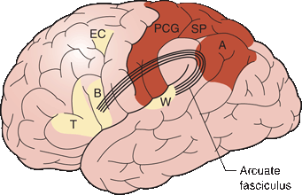
FIGURE 9.1 Centers important in language. A. Angular gyrus. B. Broca’s area. EC, Exner’s writing center; SP, Superior parietal lobule, which with the PCG (postcentral gyrus) is important in tactile recognition; T, Pars triangularis; W, Wernicke’s area.
Wernicke’s speech area lies in the superior temporal gyrus. It is essentially the sensory association cortex that lies just posterior to the primary auditory cortex. The arcuate fasciculus (AF) is a deep white matter tract that arches from Wernicke’s area around the posterior end of the Sylvian fissure and through the subcortical white matter of the insula to Broca’s area. Other tracts in the subcortical white matter of the insula provide additional connections between the PIF and PST areas. Some have suggested the AF actually connects with Broca’s area through a relay station in the premotor/motor areas. The angular gyrus is part of the inferior parietal lobule; it caps the posterior ramus of the Sylvian fissure and lies between Wernicke’s area and the visual cortex. The angular gyrus is important for reading and similar nonverbal language functions. The supramarginal gyrus also lies between the visual cortex and the posterior perisylvian language areas and is involved with visual language functions. Exner’s center is a purported cortical area concerned with writing that lies in the middle frontal gyrus of the language-dominant frontal lobe very near the frontal eye field, just anterior to the primary motor cortex for the hand. There may be white matter tracts connecting Wernicke’s and Exner’s areas analogous to the AF.
Although the cortical areas and connections described above are important language centers, the clinicopathologic correlations are not so exact as to permit precise localization in all instances. The degree of deficit seems to correlate with the size of the lesion as well as its location. Language functions are not as discretely localized in the brain as are things such as vision and elemental sensation, but they are more localized than such faculties as intelligence, judgment, and creativity. There is evidence that propositional speech depends on left hemisphere regions remote from the classic perisylvian language areas. The 2012 meta-analysis of more than 100 functional imaging studies done by Dewitt and Rauschecker implicates a much broader portion of the superior temporal gyrus in speech comprehension than has been previously appreciated, challenging the classical scheme that word recognition occurs in the PST gyrus. These studies show that activation associated with the processing of phonemes localizes to the midportion and the processing of words to the anterior portion of the superior temporal gyrus. The perisylvian language areas are perfused by the middle cerebral artery (MCA); the anterior language areas are supplied by the superior division and the posterior areas by the inferior division. Most cases of aphasia are due to ischemia in the MCA distribution.
EXAMINATION OF THE PATIENT WITH APHASIA
Initial appraisal of language function takes place during the taking of the history. Obvious deficits require exploration, but there may be language deficits that are not readily apparent during history taking. For example, the inability to repeat, which is the essential characteristic of conduction aphasia, may not be apparent during history taking. Some degree of formal assessment is usually prudent. In evaluating aphasia, it is important to know about the patient’s handedness (and sometimes the familial tendencies toward handedness), cultural background, native language and other languages spoken, vocabulary, educational level, intellectual capacity, and vocation. Just as it is not possible to evaluate mental status in a patient with impaired language function, it is difficult to evaluate language status in a person who has altered mental status causing confusion, disorientation, inattention, agitation, or severe depression, or who is emotionally distraught. Patients with emotional problems may have language disturbances on a nonorganic basis. Any apparent speech or language difficulty must of course be correlated with the findings on other parts of the neurologic examination.
About 90% to 95% of the population is right-handed. The left cerebral hemisphere is dominant for language in 99% of right handers, and 60% to 70% of left handers. Of the remaining left handers, about half are right hemisphere dominant and about half have mixed dominance. Shifted sinistrals (anomalous dextrals) are naturally left-handed individuals forced by parents or teachers early in life to function right handed, primarily for writing. This approach to dealing with left handedness has largely died out, but shifted sinistrals are still encountered, primarily in the older population. One can therefore encounter right-handed patients (dextrals) who are left-hemisphere dominant for language, left-handed patients (sinistrals) who are still left-hemisphere dominant, “righthanded” patients who are right-hemisphere dominant (anomalous dextrals), and left-handed patients who are right-hemisphere dominant (true sinistrals). Since clinical abnormalities of higher cortical function, especially language, are heavily influenced by dominance, determination of the patient’s handedness and dominance status is paramount. Only about 2% of cases of aphasia are due to unilateral right hemisphere lesions.
Cerebral dominance and handedness are at least in part hereditary. Failure to develop clear hemispheric dominance has been offered as an explanation for such things as dyslexia, stuttering, mirror writing, learning disability, and general clumsiness. Many patients are at least to some degree ambidextrous, and it may be difficult, short of a Wada test, to be certain which hemisphere is language dominant. Various “foolproof” markers of true handedness have been proposed but all are suspect. In right-handed patients, aphasia will be due to a left hemisphere lesion in 99% of the cases; the other 1% are crossed aphasics. In left handers, the situation is much more variable. In one series of left-handed aphasics, 60% had lesions of the left hemisphere. There may be a degree of mixed dominance for language in non–right-handed individuals. Aphasia may tend to be less severe in left handers and recover better; just a family history of left handedness in a right-handed aphasic may predict better recovery. Basso has challenged the concept of better recovery in non–right-handed patients.
Multilingual aphasics require examination in all of their languages. Polyglots may have several centers for speech in somewhat discrete but overlapping cortical areas. Neurophysiologic and neuroimaging studies are gradually adding to our knowledge of the regions of the brain involved in the various speech-related processes. In bilinguals, the cerebral representation of some functions is similar for both languages, but the areas concerned with other functions may be different depending on when the languages were acquired. Which language recovers best in multilingual aphasics is variable. Pitres’ law states that recovery from aphasia will be best for the language most used, but Ribot’s rule holds that recovery will be best for the native language. In fact, most patients show parallel recovery in both languages.
There are six separate components of language function that are typically tested in the clinical arena: spontaneous (conversational) speech, auditory comprehension, naming, reading, writing, and the ability to repeat. It is often useful to assess these components individually before trying to synthesize the findings into a diagnostic entity. There are several instruments available for more detailed examination of the aphasic patient, such as the Boston Diagnostic Aphasia Examination, Western Aphasia Battery, and others. The Western Aphasia Battery produces a summary score reflecting overall severity (aphasia quotient). For clinical purposes, it is not clear these add a great deal to the bedside examination.
SPONTANEOUS SPEECH
In addition to high-level propositional speech, spontaneous utterances may include the lower-level functions of emotional and automatic speech. Emotional speech is spontaneous speech prompted by a high emotional charge. It is present in animals, especially higher primates, and in humans before they acquire propositional language. Some patients with aphasia, primarily nonfluent aphasia, even when severe, may swear and curse eloquently when angry, often to the shock and surprise of friends and family. Automatic speech refers to the recitation of simple overlearned items from early childhood or to a specific retained speech fragment that an aphasic patient is still capable of saying even in the presence of severe nonfluency. Even when unable to produce propositional speech, an aphasic patient may be able to automatically count, say the days of the week or months of the year, repeat the alphabet, say his name, or recite nursery rhymes. Some aphasic patients are able to sing simple overlearned songs, such as Happy Birthday, even when they are unable to speak.
A retained fragment that an aphasic patient repeats over and over has been referred to as a monophasia (recurring utterance, verbal stereotypy, verbal automatism, verbigeration). In monophasia, the individual’s vocabulary is limited to a single word, phrase or sentence, such as “do-do-do” or “Oh, God.” Verbal automatisms occur most often in global aphasia. The recurrent utterance may be a real word or a neologism. Sometimes the monophasia is an outrageous expletive that bursts from an otherwise dignified and respectable patient under socially awkward circumstances. Some verbal automatisms are unusual and difficult to understand. One aphasic patient would say “Pontius Pilate” in response to any and all questions. Other examples include, “no pasta,” “television,” and “gotta go.” Broca’s original aphasic patient, M. Leborgne, was nicknamed “Tan” because that was the only word he could say. According to Critchley, Hughlings Jackson first became interested in aphasia when his family vacationed in a house where the aphasic landlady could utter only the neologistic stereotypy “watty.” A patient may have several stereotypies in their repertoire, and preservation of stereotypic social responses (“hello,” “fine”) may trick the careless or rushed clinician into believing the patient is linguistically intact. Speech automatisms can also occur as an ictal phenomenon.
A paraphasia is a speech error in which the patient substitutes a wrong word or sound for the intended word or sound. Paraphasic errors are common in aphasic patients. In a phonemic (phonologic, literal) paraphasia, there is the addition, deletion, or substitution of a phoneme; however, the word is recognizable and may be clearly pronounced. Substitution of the wrong phoneme would cause the patient to say “blotch” instead of watch, or “thumbness” instead of numbness. Technically, a literal paraphasia is a single-letter substitution. Phonemic paraphasia is the preferable term since a single letter substitution also changes the phoneme, and the brain thinks in phonemes, not letters. Illiterate patients commit phonemic paraphasias despite their ignorance of letters. In a semantic (verbal) paraphasia, the patient substitutes the wrong word. A semantic paraphasia would cause the patient to say “ring” instead of watch. Paraphasias are similar to the malapropisms, spoonerisms, and sniglets everyone occasionally utters, but aphasic patients make them more often and may not recognize them as wrong. A neologism is a novel utterance, a nonword made up on the spot. The patient might call a watch a woshap. Phonemic paraphasias are more typical of anterior, and semantic paraphasias more typical of posterior, perisylvian lesions.
In evaluating propositional speech, note pronunciation, word and sentence formation, fluency, cadence, rhythm, prosody, omission or transposition of syllables or words, misuse of words, circumlocutions, repetition, perseveration, paraphasias, jargon, and the use of neologisms. Aphasic patients may use unusual synonyms or circumlocutions in order to avoid the use of a word that cannot be recalled. There may be omissions of words; hesitations and inappropriate pauses; perseveration; difficulty understanding the implication of words; verbal automatisms; agrammatism; jargon or gibberish. When the patient is having difficulty with fluency, it is difficult to evaluate propositional spontaneous speech. Fluency refers to the volume of speech output. Normal speech is 100 to 115 words per minute. Speech output is often as low as 10 to 15 words per minute, sometimes less, in patients with nonfluent aphasia. If the maximum sentence length is fewer than seven words, then the patient is nonfluent. Patients are usually aware of nonfluency and frustrated by it. Their speech may tend toward the laconic, answering questions but trying to speak no more than necessary. Patience and open-ended questions are the best approaches in persuading the patient to converse. Patients unable to express themselves through speech may use pantomime or gesture, shaking or nodding the head, shrugging the shoulders, or demonstrating visible emotional reactions. In severe aphasia, the patient may be unable to utter a single word.
COMPREHENSION
The patient’s responses to verbal requests and commands and to everyday questions and comments give information about his ability to understand speech. Comprehension may be tested by having the patient follow verbal commands (“show me your teeth,” “stick out your tongue,” “close your eyes,” or “point to the ceiling”). Comprehension can be judged to be reasonably intact if the patient follows a complicated, multistep command. However, failure to follow a command, even a simple one, does not necessarily prove that comprehension is impaired. A patient may not comply because of apraxia. Patients with a left hemisphere lesion may even have apraxia for functions of their nonparetic left hand. They may be unable to salute, wave goodbye, or perform other simple functions on command using the left hand because of involvement of fibers that transmit information from the language areas on the left to the motor areas on the right (sympathetic apraxia). When the patient does not follow simple commands, establish whether he can say or shake his head yes and no. Then ask ridiculously simple questions, such as—”Are you from the planet Jupiter?”, “Did you have nails for breakfast?”, “Are you riding in a taxicab?”, or “Are you a man (or a woman)?” The responses may be nonverbal. An elderly woman who laughs when asked “Are you pregnant?” has understood the question. More complex yes-no questions might include the following: “Is a mother older than her daughter?”, “Do you have dinner before breakfast?”, “Can you fly in a car?”, “Did the sun come up this morning?”, or “Do you have feet on the ends of your legs?” Because the chance of a correct response is 50%, it is important to ask enough questions to exclude lucky answers.
Impaired comprehension may result from difficulty understanding grammar and syntax, words in relation to other words, or difficulty with semantics, understanding individual words. The patient may have more difficulty with polysyllabic words and long sentences than with simple words and short sentences. Compound sentences and double or complex commands may be used to see if comprehension is more than superficial. The aphasia examination begins to overlap with the mental status examination with commands such as “place one coin on the table, give me the second, and keep the third in your hand” or “here is a piece of paper; tear it in four parts and place one on the table, give one to me, and keep two for yourself” (Marie’s paper test). Both comprehension and retention are evaluated by telling a short story and then asking questions about it. Patients with impaired comprehension have particular difficulty with passive constructions (e.g., “The lion was killed by the tiger; which animal is dead?” or “The boy was slapped by the girl; who got hit?”) and possessives (e.g., “Is my wife’s brother a man or a woman?”). Patients who are unable to comprehend spoken or written language may understand pantomime, gestures, and symbols. They may imitate the examiner in placing a finger to the nose or sticking out the tongue. Imitation, however, is a more lower-level function than comprehension.
Many aphasic patients have difficulty with right-left orientation, especially with posterior lesions. Right-left confusion is part of Gerstmann’s syndrome. Testing right-left orientation might include such commands as “show me your right thumb” or “touch your right ear with your left thumb.” It is important to determine baseline function before concluding a patient has right-left confusion.
NAMING
Testing naming ability is an important part of the aphasia examination. Naming is a delicate function, and most aphasic patients have some difficulty with it. However, naming defects are nonspecific. In anomic aphasia, an inability to name is an isolated defect, but more often misnaming occurs as part of some other aphasic, or even nonaphasic, syndrome. In confrontation naming, the patient is asked to name simple objects such as a key, pencil, coin, watch, parts of the body (nose, ear, chin, fingernail, knuckle), or to name colors. When lost for the name of an object, the patient may describe it or tell its use. The patient may be able to name an object, such as a watch, but be unable to identify the component parts, such as the band or buckle. Some caution is necessary, as there are age, cultural, and even gender influences at work. For whatever reason, many normal women are unable to identify a watch crystal. Many normal men (with intact color vision) are unable to name more than primary and very simple secondary colors. Before including something as a naming test item, the examiner should ensure that nonaphasic people of all ages and both sexes are normally able to identify it. Some normal patients use unusual names for various parts of the body, especially the fingers. Some of this is related to educational level and region of origin. Individuals may refer to the index finger or pointer as “the finger next to the thumb,” or call it the “dog finger,” “poison finger,” or “statue of liberty finger.” These patients are not aphasic. Many normal individuals cannot name the index, middle, and ring fingers. When unable to retrieve a name, an aphasic patient may be able to select the correct name from a list. Another naming test is to have the patient point to something named by the examiner (e.g., the telephone, the window).
A sensitive method of testing spontaneous naming ability is word list generation. The patient is asked to name as many items as possible in a certain category in 1 minute. Animals are a common category for testing spontaneous naming. The patient may name any types of animals (e.g., farm, zoo, etc.), but groups should not be suggested ahead of time since there may be an inability to shift groups. It is wise to check more than one item category; other useful categories include tools, foods, countries, and modes of transportation. Spontaneous naming ability also depends on age and educational level. Normal patients should name a minimum of 12 items in a category; some adjustment may be necessary for poorly educated and older patients. Another measure of spontaneous naming is to ask the patient to list all of the words he can think of that begin with a certain letter. The FAS test is popular. The patient thinks of words beginning with one of these letters, excluding proper nouns or morphological variants. For FAS, a person of average education should produce 12 or more words per letter in 1 minute, or 36 words with all three letters in 3 minutes. Standardization and reference values for testing naming are imperfect. Language competence depends on education, dialect, experience, and other factors. Often the reference population does not include less well-educated people, nor every dialect. Poor word list generation may also occur with dementia, depression, parkinsonism, and prefrontal lesions. Responsive naming is also useful, and uses audition rather than vision. The patient may be asked for nouns (e.g., “Where do teachers work?”), verbs (e.g., “What do you do with a cup?”), or adjectives (e.g., “How does sugar taste?”).
REPETITION
The ability to repeat may be selectively involved or paradoxically preserved in certain aphasic syndromes. Most often the inability to repeat is proportional to the defect in comprehension or fluency, and repetition is a good screening test for aphasia. The patient is asked to repeat words or phrases back to the examiner. A patient’s repetition span (i.e., the number of words he can repeat) is usually two more than his digit span. Simple repetition tasks might include counting, avoiding numbers that might be repeated by automatic speech, or repeating single words. More complex tasks include polysyllabic words (e.g., catastrophe), phrases (e.g., “If he were here, I would go away”), or tongue twisters (e.g., Popocatepetl [po-pócah-té-petl], a volcano in Mexico). The stock phrases used to test for dysarthria work for this purpose as well. A popular phrase for testing repetition in aphasia is “no ifs, ands, or buts.” Omitting the s in each of these words may not be an error in some dialects of English. A better repetition test is “they heard him speak on the radio last night’ (modified from the Boston diagnostic aphasia examination). Patients with impaired repetition may omit words, change the word order, or commit paraphasic errors. Repetition is preserved in anomic, transcortical, and some cases of subcortical aphasia.
WRITING
The patient’s ability to use written language should also be assessed. It may be disturbed in conjunction with abnormalities of spoken language, or separately. Patients who are aphasic in speech are also aphasic in writing, but writing may be preserved in patients with dysarthria or verbal apraxia. In all aphasias, reading and writing are typically worse than understanding and speaking, probably because they are secondarily acquired skills. The patient may be asked to write spontaneously or to dictation. A spontaneous writing sample might include a few words, a sentence, or a paragraph. The writing sample usually reveals the same sorts of naming difficulties and paraphasias evident in the patient’s speech. Patients may be able to write elementary, overlearned things such as name, address, days of the week and months of the year, but be unable to write more complex material. There may be a difference in the patient’s ability to print and to write in cursive. The ability to write to dictation is analogous to the ability to repeat verbal material. Copying written material also assesses the ability to transfer information from the visual system to the language areas. Having the patient copy written material may also test the connections between the receptive language areas and Exner’s writing center. However, copying does not require much processing; one can copy material in another language despite not speaking the language, as long as the alphabet is the same. An inability to copy may be due to apraxia. Naming can also be tested by having the patient write down the names of things in a manner similar to that for speech.
READING
The patient’s ability to comprehend written language symbols can be tested by having him read. Written language is perceived by the visual system and the information conveyed to the perisylvian language centers. Dysfunction of the language centers or interruption of the connections with the visual system may cause an inability to read (alexia). Reading difficulty due to acquired alexia is unrelated to the developmental (congenital) dyslexia seen most often in school-age boys that may cause severe reading disability. Patients may have alexia without any accompanying inability to comprehend speech—the syndrome of pure word blindness. Alexia may occur with or without a hemianopia. Alexia may occur with or without accompanying agraphia. Most patients with alexia also have difficulty with writing (alexia with agraphia). Some patients have alexia without agraphia (see Chapter 10). Judging reading ability by having the patient follow a written command such as close your eyes involves a praxis element and should be interpreted with caution. For patients unable to read aloud, use questions that can be answered by “yes” or “no,” or by gestures. It is also important to determine whether the patient is able to read his own writing.
Reading aloud is a different task from reading comprehension. Oral reading (visual input-oral output) is comparable to copying (visual input-manual output), repetition (auditory input-oral output), and transcribing dictation (auditory input-manual output), and may be preserved despite impaired reading comprehension.
CLASSIFICATION OF THE APHASIAS
Classification of the aphasias is problematic. These disorders vary in severity, even with a lesion in the same location, and are frequently mixed in type. There have been many attempts at classification from anatomic, physiologic, and psychological points of view. None is entirely satisfactory. A strictly anatomic classification does not apply in all instances, for a small lesion may cause severe impairment of both fluency and comprehension, while an extensive lesion sometimes causes an isolated defect. Lesions similar in size and location on imaging studies may be associated with different aphasic syndromes even in persons with identical cerebral dominance for speech. Lesions in different locations and of variable size may produce similar aphasic syndromes. Nevertheless, some general relationships exist between anatomic sites and the type of aphasia.
One common classification divides aphasias into expressive and receptive types. In expressive aphasia, the patient has difficulty with speech output and struggles to talk (nonfluent); in receptive aphasia, the primary difficulty is with understanding language, while speech output is unaffected (fluent). A major problem with the expressive-receptive classification of aphasia is that all aphasic patients have difficulty expressing themselves. This causes difficulty, particularly for trainees and nonneurologists. There is a tendency to classify almost all aphasias as expressive, even when they are flagrantly receptive. It requires some clinical experience to recognize that a patient may be having difficulty expressing himself linguistically because of a defect in the reception (comprehension) of spoken language. Other simple dichotomous classifications proposed include: fluent/nonfluent, motor/sensory, and anterior/posterior. Although each of these is useful, none adequately describes most aphasic patients, who have some evidence of both types. Pure forms of aphasia are uncommon; most patients with sensory aphasia have some motor deficit, posterior lesions can cause nonfluency, anterior lesions can cause comprehension deficits, and aphasic disorders can occur with pathology that does not directly affect the classical perisylvian language centers, such as subcortical and even nondominant hemisphere lesions.
The Wernicke-Geschwind model (Boston classification) recognizes eight aphasia syndromes: Broca’s, Wernicke’s, conduction, global, transcortical motor, transcortical sensory, transcortical mixed (isolation of the speech area), and anomic. It divides aphasias into fluent and nonfluent varieties (Tables 9.2 and 9.3). If speech output is high and articulation facile, the aphasia is referred to as fluent; if speech output is sparse and effortful the aphasia is classified as nonfluent. Nonfluency occurs when a lesion involves the anterior speech areas in the region of Broca’s area in the frontal lobe. When these areas are relatively spared, fluency is preserved. Broca’s is a type of nonfluent aphasia.
TABLE 9.2 The Major Aphasia Syndromes
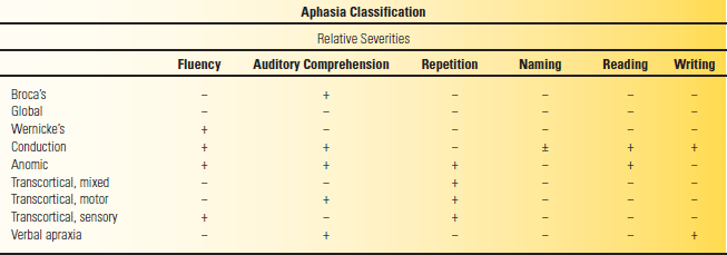
+, function is relatively intact; −, function is abnormal; ±, involvement is mild or impairment equivocal. Modified from Campbell WW, Pridgeon RP. Practical Primer of Clinical Neurology. Philadelphia: Lippincott Williams & Wilkins, 2002.


