, Nima Etminan1 and Daniel Hänggi1, 2
(1)
Neurochirurgische Klinik, Universitätsklinikum Düsseldorf, Düsseldorf, Germany
(2)
Medical Art Christine Opfermann-Rüngeler, Zentrum für Anatomie Heinrich Heine Universität, Düsseldorf, Germany
5.1 Approach to the Aneurysm
When operating early after subarachnoid hemorrhage, the anatomy may be much obscured by the cisternal and superficial blood accumulation and by the reactive swelling. It is of utmost importance to ensure sufficient brain relaxation prior to opening the dura. The ventricular or spinal CSF drain should be opened following craniotomy and 50–100 mL of CSF should be drained. It is important not to open the dura while it is still under tension, because doing so could result in brain prolapse and premature rerupture of the aneurysm. At this point, it is also important to check arterial pressure and communicate with the anesthesiologist. We recommend keeping the arterial pressure at a level corresponding to the preoperative value minus the preoperatively measured intracranial pressure (ICP). If preoperative ICP levels are not available, 10 mmHg is used as an average estimate, and arterial pressure during surgery should therefore be maintained at 10 mmHg lower than the preoperative value. This directive keeps the cerebral perfusion pressure steady, thus both avoiding ischemia and controlling the risk of premature rerupture of the aneurysm.
5.2 Opening the Cisterns
Once the dura has been opened, the aim is to gain proximal control and free the neck of the aneurysm with as little dissection as possible. Any dissection in the vicinity of the aneurysm carries the risk of rupture. The only location where the fragile aneurysm dome is not encountered with certainty is the parent artery. Therefore, the parent artery should be identified, and dissection to the aneurysm neck should strictly adhere to this vessel. The parent artery is our friend.
5.3 Gaining Proximal Control
The method of gaining proximal control depends essentially on the specific aneurysm location. It is generally not necessary to dissect the entire parent artery beginning with its origin from the internal carotid artery. In the case of an anterior communicating artery aneurysm, it is sufficient to look for the A1 in its middle segment, at the level where it crosses the optic nerve. In the case of a middle cerebral artery aneurysm (particularly when using the smaller sylvian approach), the M2 segments are usually identified first in the depth of the sylvian fissure, and these are followed backward to the bifurcation in order to secure inflow from M1 and the aneurysm neck.
Gaining proximal control always provides only relative safety. Complete control of aneurysm inflow, which is necessary to deal with complex aneurysms, can be achieved only by controlling all afferent and efferent arteries. In the case of anterior communicating artery aneurysms, for example, both A1 and A2 segments must be controlled—keeping in mind that sometimes unexpected arteries can be present, such as a third A2 segment.
For noncomplex aneurysms, gaining proximal control is less important than achieving complete understanding of the anatomy of the perianeurysmal vascular complex. Therefore, no efforts should be made to control a contralateral nondominant A1 hiding behind a downward-projecting aneurysm at the anterior communicating artery, for example.
5.4 Dissecting the Neck of the Aneurysm
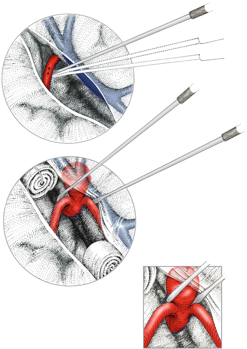
Having gained access to the parent artery, the neck of the aneurysm is approached along this artery. At this point, the importance of a clear mental image of the expected configuration of the neck in relation to the parent and branching arteries cannot be overemphasized. It is therefore important to study the preoperative 3D reconstructions of the angiography exactly, also from the direction of approach. Nonetheless, details may not be evident from the angiography, so knowledge of the common variations is important. After the widening of the parent artery suggesting that the aneurysmal neck has been defined (Fig. 5.1), both sides of the neck must be separated from the arterial branches sufficiently to allow the smooth passage later of the clip blades (in general, at least 3 mm). The dome of the aneurysm is left untouched at this stage. Separation of the neck from the arteries needs to be complete so that the dissector can be passed through.


Fig. 5.1
Following identification of the proximal parent artery or a distal branch, this artery is followed to the aneurysm-bearing bifurcation and the aneurysm neck. The neck is subsequently freed on both sides, while leaving the aneurysm dome untouched
Because the back side of the neck is not visible, a common error is to assume that it looks the same as the visible front side, but this may not be the case. The neck may be directed backward from the line of sight, thus bulging on the back side. The other variant, that the aneurysm is projecting somewhat toward the surgeon, is usually more easily realized.
5.5 Temporary Clipping, Pharmacological Neuroprotection, and Induced Hypotension
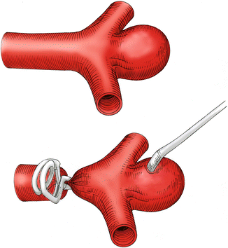
Temporary clipping has proven to be a very useful adjunct for aneurysm clipping [1]. We recommend the general use of proximal temporary clipping whenever the neck of the aneurysm is wider than the parent artery (Fig. 5.2). We do not recommend the use of temporary clipping during dissection as it usually takes considerable time and its benefit is questionable, in our opinion.


Fig. 5.2


Primary proximal temporary clipping is recommended whenever the diameter of the aneurysm neck exceeds the physiological diameter of the parent artery. Sufficient decompression of the aneurysm must be confirmed by palpation prior to proceeding further
Stay updated, free articles. Join our Telegram channel

Full access? Get Clinical Tree


