DYSTONIA

CHAPTER 35 DYSTONIA


The dystonias are an unusual group of movement disorders whose main feature is involuntary muscle contraction or spasm. The term dystonia was originally introduced by Hermann Oppenheim in 1911 to describe alterations in muscle tone and postural abnormalities that are seen in this condition. The concept of dystonia can be confusing because the term has been used to describe a symptom (e.g., a dystonic arm posture), a disease (primary torsion dystonia), or a syndrome. The dystonias constitute a relatively common group of movement disorders that encompass a wide range of conditions from those in which the only manifestation is dystonic muscle spasms to those in which dystonia is one part of a more severe neurological condition.


DEFINITION AND CLASSIFICATION


Dystonia is characterized by involuntary sustained muscle contractions affecting one or more sites of the body, frequently causing twisting and repetitive movements or abnormal postures.1 The movements range from slower twisting athetosis to rapid, shocklike jerky movements. They are repetitive and sometimes rhythmic and can be accompanied by tremor. Dystonic movements can be aggravated by movement (action dystonia) that can be nonspecific or task-specific (e.g., writing). Over time the dystonia can occur with less specific movements and eventually can be present at rest, leading to sustained abnormal postures.


Three basic approaches are used to classify dystonia: age at onset, distribution of affected body parts, and etiology. The categories of age at onset and affected body distribution (Table 35-1) are important in describing clinical signs and have clinical implications for prognosis and treatment.


TABLE 35-1 Classification of Dystonia


±, with or without.


EPIDEMIOLOGY


The true population incidence and prevalence of dystonia are unknown. The prevalence data available are usually based on studies of diagnosed cases only and therefore are underestimates of the real numbers. This is particularly the case with dystonia, which can manifest in a variety of ways, and a significant number of cases of focal dystonia are undiagnosed or even misdiagnosed. In an early study in the United States that was based on case note review, the prevalence for PTD was estimated to be 329 per million population. In more recent studies of diagnosed cases in Japan and Europe, the prevalence was estimated to be between 101 and 150 per million. The most reliable estimate is from an ongoing study in the northeast of England, where ascertainment was more complete, and some previously undiagnosed cases were identified. This finding has implied a prevalence rate of 485 per million. The prevalence of secondary dystonia is unknown, although it is estimated from case series that it may be less than 20% to 25% the rate for PTD.


The most prevalent form of PTD is focal dystonia, of which cervical dystonia is the commonest, with prevalence rates reported between 57 and 290 per million population. Rates for blepharospasm are 17 to 80 per million, and for writer’s cramp, 14 to 61 per million.


In a study in South Tyrol in Austria, a random sample of the population older than 50 years was examined.2 Primary dystonia was diagnosed in 6 of the 707 individuals studied, implying a prevalence of 7320 per million in this age-selected population, although 95% confidence intervals were very wide, at 3190 to 15,640, because of the small sample. However, this indicates that in the aging population, dystonia is a relatively common neurological disorder.


CLINICAL FEATURES


The diagnosis of dystonia is based on clinical findings. Primary dystonia has no other neurological features apart from dystonia and tremor.3 Features suggestive of a secondary cause of dystonia are listed in Table 35-2. Investigations are usually performed to help rule out a secondary cause of dystonia.


TABLE 35-2 Clinical Features Suggestive of Secondary Dystonia


Primary Dystonia


Early-Onset Primary Torsion Dystonia (Dystonia Musculorum Deformans, Oppenheim’s Dystonia)


The commonest cause of early onset PTD is mutation in the DYT1 gene on chromosome 9q34, which is inherited as an autosomal dominant trait with reduced penetrance (30% to 40%). The disorder typically manifests in childhood or adolescence (mean age at onset, 12 years) with dystonia causing posturing of a foot, leg, or arm. Dystonia is usually first apparent with specific actions (e.g., writing or walking) but becomes evident with less specific actions over time and spreads to other body regions. No other neurological abnormalities are present apart from postural arm tremor. Disease severity varies considerably even within the same family, and isolated writer’s cramp may be the only sign. However, approximately 60% to 70% of individuals have progression to generalized or multifocal dystonia involving at least a leg and an arm, and often axial muscles. In 10% of cases, segmental dystonia develops, and only 25% remain focal. The cranial muscles are involved in about 10% of affected patients.


Key investigations are to exclude treatable differential diagnoses, such as Wilson’s disease and dopa-responsive dystonia (DRD). DYT1 dystonia is diagnosed through molecular genetic testing of the TOR1A gene, which reveals a three-base pair deletion in all affected individuals. There is a higher prevalence of DYT1 PTD in the Ashkenazi Jewish population, which is the result of a founder mutation that appeared about 250 years ago.


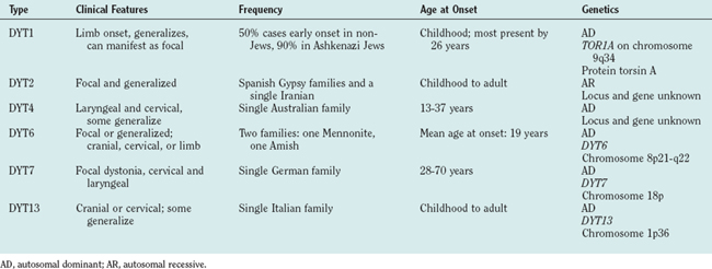
Most forms of early-onset PTD are genetic in origin, and Table 35-3 lists the genetic forms that have been identified to date. Most are autosomal dominant, some reported only in single families. The existence of autosomal recessive forms (DYT2) is controversial.


Focal Primary Torsion Dystonia


These are by far the commonest forms of dystonia. Usually sporadic, they have onset in adult life and remain focal in distribution. Families with autosomal dominant forms have been described, and it is believed that a proportion of the apparently sporadic cases may represent manifestation of a dominant gene with very low penetrance (estimated at 12% to 15%). The individual types are discussed as follows.


Cervical dystonia (spasmodic torticollis)


Cervical dystonia is a focal dystonia that affects cervical muscles, leading to abnormal postures and movements of the head, neck, and shoulders. It is the most common form of dystonia, usually with onset in the fifth decade (mean age at onset, 42 years) and affects women more than men (ratio, 1.4 : 1 to 1.6 : 1). The dystonic muscle activity can be tonic, phasic, or tremulous and leads to symptoms of neck pain, head posturing, or repetitive jerking, producing tremor of the head. Cervical dystonia symptoms tend to worsen over the first 5 years and then stabilize. Twisting of the head around the horizontal axis (torticollis) is the most common movement, present in 80% of patients and caused by overactive contralateral sternomastoid and ipsilateral splenius capitis muscles. Laterocollis (head sideways) is seen in 10% to 20% and caused by overactivity in the ipsilateral splenius, sternomastoid, and levator scapulae muscles. Retrocollis (head back) and antecollis (head forward) are less frequent. Many patients, however, present with combinations of torticollis and laterocollis. Pain is present in 75% of patients and can cause significant disability.


Many patients with cervical dystonia have sensory tricks (geste antagoniste) that can alleviate symptoms. These can involve touching the back of the head, cheek, or temple and lead to reduction in abnormal dystonic muscle spasm. Spontaneous remission of symptoms occurs in less than 20% of patients; unfortunately for most of these patients, there will be a subsequent relapse. Focal cervical dystonia can spread to other body parts, including the face and arms, but rarely generalizes.


The long-term complications of cervical dystonia include cervical spine degeneration, which leads to radicular or myelopathic symptoms. Cervical dystonia also has a significant effect on quality of life and is associated with a higher incidence of anxiety and depression.


Writer’s cramp and task-specific limb dystonias


Writer’s cramp is the most common form of task-specific dystonia and, in contrast to craniocervical dystonia, is more common in men than in women. Onset usually occurs between the ages of 30 and 50 years and often starts with a feeling of tension in fingers and forearms that interferes with writing fluency. The pen is held abnormally forcefully as a result of dystonic contraction of hand and/or forearm muscles. This commonly involves excessive flexion of the thumb and index finger with pronation of the hand and ulnar deviation of the wrist. Affected individuals may also experience lifting of the thumb or index finger off the pen or isolated extension of fingers. Up to 50% of patients also experience upper limb tremor, either on writing or a postural tremor. Strain and aching, particularly in affected forearm muscles, is common on writing, but pain is an uncommon feature.

Writing difficulty is often intermittent at onset but usually progresses so that cramping starts soon after starting writing. In a minority of patients, dystonia occurs on performing other manual tasks, and this can be a feature that develops with time. Spontaneous remission is rare. Many patients whose writing has become illegible therefore learn to write with their nondominant hands. Unfortunately, in up to 10% of cases, writer’s cramp can develop in that hand as well.


Dystonic patterns of involuntary muscle contractions are also seen in the context of other highly learned motor skills. These are most commonly seen in professional musicians, craftsmen, and sportsmen whose work involves frequent, repetitive movements of particular muscle groups. They have been reported in fewer than 1% of professional musicians, more frequently men. In pianists, for instance, the fourth and fifth fingers of the right hand are most commonly involved, whereas for guitarists, the third finger of the right hand is affected. For wind instrument players, the hand supporting the instrument and doing fingering at the same time is most involved. Less commonly, lip or orofacial dystonia can develop. Tremor can accompany task-specific dystonias in less than 40% of cases. Other manual tasks associated with task-specific dystonia include typing, painting, and sports such as golf, tennis, and snooker.


Laryngeal dystonia


Laryngeal dystonia is relatively rare and can be divided into cases in which the predominant problem is spasm of the adductor muscles and spasms involving abductor muscles. It can occur in isolation or sometimes as part of a segmental craniocervical dystonia. In one family with autosomal dominant inheritance (DYT3 locus), it is the presenting feature in individuals who developed generalized dystonia.


Adductor spasmodic dysphonia is the commonest form and is characterized by intermittent voice stoppages, particularly with vowels. This is caused by hyperadduction of the vocal cords caused by involuntary spasm of thyroarytenoid and/or lateral cricoarytenoid muscles. Abductor spasmodic dysphonia occurs in about 15% of patients and manifests with breathy breaks in speech, especially with consonants. In some cases, this is caused by involvement of vocal fold opening muscles, such as the posterior cricoarytenoid and cricothyroid muscles. Spasmodic dysphonia is task specific and occurs only during speech. Laughter, crying, and breathing are unaffected. Voice tremor often occurs with both types of spasmodic dysphonia and is also speech specific.


Other laryngeal disorders that can be confused with laryngeal dystonia can occur in other neurological conditions, but the presence of additional neurological signs should alert the clinician to alternative diagnoses. Examples are vocal fold paralysis in motor neuron disease, airway obstruction in multiple-system atrophy, hypophonia in Parkinson’s disease, and abductor vocal fold paralysis in hereditary motor neuronopathy.


Secondary/Symptomatic Dystonias


Dystonia-Plus Syndromes


Dystonia-plus syndromes describe a group of conditions that can be distinguished from PTD on the basis of clinical characteristics found in addition to dystonia, or specific pharmacological responses. They usually have a genetic etiology but do not have underlying neurodegeneration. The group comprises three distinct conditions: DRD, myoclonus dystonia syndrome, and rapid-onset dystonia parkinsonism (RDP).


Dopa-responsive dystonia


DRD was first described in Japan in 1977 by Masaya Segawa. Patients typically present in childhood with gait disturbance caused by foot dystonia. The dystonia frequently worsens as the day goes on (diurnal variation) and is relieved by rest or sleep. Progression is variable; some patients develop severe generalized dystonia, whereas others develop features suggestive of lower limb spasticity. Parkinsonian features such as bradykinesia and rigidity can develop in later life in some affected individuals but can also be the presenting features in adult life in a minority of cases. On occasion, DRD can manifest with adult-onset limb dystonia (e.g., writer’s cramp), with cranial or cervical dystonia, or with signs resembling spastic paraplegia. In most cases, DRD is inherited as an autosomal dominant trait with reduced penetrance.


The key feature in DRD is a dramatic and sustained response to small doses of levodopa (L-dopa), often as low as 50 to 200 mg. Benefit is usually apparent within days to weeks, and the motor complications of L-dopa treatment seen with Parkinson’s disease rarely develop, even with long-term treatment. Anticholinergic drugs also can be beneficial.


The principal considerations in the differential diagnosis for childhood DRD are early-onset PTD, spastic paraplegia and cerebral palsy, and early-onset parkinsonism, especially when it is caused by mutations in the parkin gene. The patients thought to have early-onset parkinsonism often present with dystonia and show good initial response to L-dopa. However, clues to the diagnosis come from the inheritance pattern (usually autosomal recessive for the parkin gene) and the occurrence of motor fluctuations and dyskinesias with L-dopa treatment. Positron emission tomography (PET) with markers for presynaptic dopaminergic terminals (18F-dopa) or single photon emission computed tomography can also differentiate between the two conditions.


The gene for dominant DRD has been mapped to chromosome 14 (DYT5) and mutations within the gene for guanosine triphosphate (GTP) cyclohydrolase 1 have been identified. Numerous mutations have been identified in all five exons, which makes genetic testing laborious. Other extremely rare forms of DRD have been reported, including a recessive form with genetic deficiency of tyrosine hydroxylase and defects in other enzymes involved in pterin synthesis. The diagnosis can usually be confirmed by an excellent response to L-dopa treatment in dosages slowly increasing up to 400 mg a day. Alternatively detecting reduced levels of pterins in the cerebrospinal fluid or an abnormal oral phenylalanine loading test can substantiate the diagnosis.

< div class='tao-gold-member'>

Stay updated, free articles. Join our Telegram channel

Jun 19, 2016 | Posted by in NEUROLOGY | Comments Off on DYSTONIA

Full access? Get Clinical Tree

Get Clinical Tree app for offline access