, Damien Gervasoni1 and Catherine Vogt2
(1)
Lyon Neuroscience Research Centre CNRS UMR 5292–INSERM U 1028, Université Claude Bernard Lyon 1, Lyon, France
(2)
Université Claude Bernard Lyon 1, Lyon, France
2.1 General Organization of the Nervous System
The nervous system is the control center for communication between different organs in the body. The cells of the nervous system communicate via specific electrical signals that cause rapid responses by acting on muscle or glandular effectors. The nervous system receives sensory information in the form of stimuli processed by the sensory receptors and adapts the appropriate actions to take in response to this stimulation. The nervous system acts as the headquarters of management and the director of the response. When necessary, it can provide not only a motor reaction but also secretory responses through the endocrine system. The nervous system is composed of the central nervous system (CNS), which assures the integration and the regulation of the entire nervous system, and the peripheral nervous system (PNS), which allows communication between the CNS and the rest of the body.
2.1.1 The Central Nervous System (CNS)
During ontogenesis, the formation of the brain begins with the subdivision of the neural tube into three primary swellings or vesicles (Fig. 2.1a): at the front, the prosencephalon containing the visual and olfactive centers; in the middle, the mesencephalon; and, between the mesencephalon and the spinal cord, the rhombencephalon, containing the centers of auditory function. The prosencephalon divides further into two parts (Fig. 2.1b): the telencephalon, which will evolve into the two cerebral hemispheres, and the diencephalon wherein visual information is processed by the thalamus. The rhombencephalon subdivides into the metencephalon and myelencephalon (Fig. 2.2). Each of these five secondary embryonic vesicles generates the formation of highly specialized structures of the vertebrate nervous system.
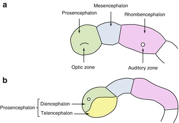
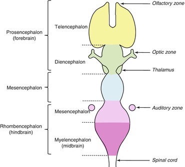
Fig. 2.1
Schematic representation of the three primary vesicles (a) and the subdivision of the prosencephalon (b)
Fig. 2.2
Schematic representation of the five secondary vesicles, the main subdivisions of the embryonic vertebrate brain
After the formation of the two hemispheres, another pair of vesicles develops on their ventral surface to form the olfactory bulbs and other structures that contribute to olfaction. Diverse structures subsequently emerge from the walls of the telencephalon at the same time as the development of the white matter that connects them. The neurons of the telencephalic wall proliferate to form three regions: the cerebral cortex, the basal forebrain, and the olfactory bulbs (Saladin 2012; Mtui and Gruener 2006; Jessell et al. 2000; Duester 2008; Dono 2003). At this point in its evolution, the neuraxis reveals two principle and distinct elements: the brain situated within the cranial vault and the spinal cord within the vertebral canal (Table 2.1).
Table 2.1
Anatomical divisions of the central nervous system
Central nervous system | Brain | Prosencephalon | Telencephalon | Rhinencephalon, amygdala, hippocampus, neocortex, basal ganglia, lateral ventricles | |
Diencephalon | Epithalamus, thalamus, hypothalamus, subthalamus, pituitary gland, pineal gland, third ventricle | ||||
Brain stem | Mesencephalon | Tecum, cerebral peduncle, pretectum, mesencephalic duct | |||
Rhombencephalon | Metencephalon | Pons, cerebellum | |||
Myelencephalon | Medulla oblongata | ||||
Spinal cord | |||||
The brain is responsible for the emission of nervous impulses to the rest of the body, the processing and integration of information originating from sensory nerves, and the more highly evolved operations that allow the association of this information so that the organism can respond appropriately. The brain is divided into four parts (Figs. 2.3, 2.4 and Table 2.1): the telencephalon or cerebrum, with its two large hemispheres (right and left) linked by the corpus callosum; the diencephalon containing the hypothalamus and thalamus; the mesencephalon or midbrain; and the rhombencephalon or hindbrain. The telencephalon and diencephalon together form the so-called forebrain. The mesencephalon (or midbrain) and rhombencephalon (or hindbrain, containing the pons and medulla oblongata) together make up the brain stem, which connects the rest of the brain to the cerebellum and spinal cord.
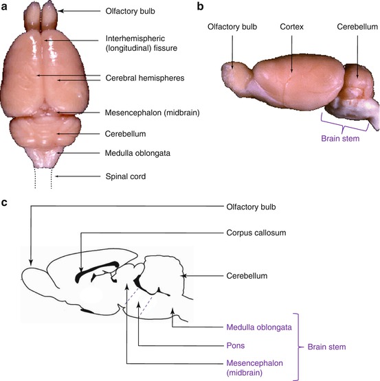
Fig. 2.3
Morphology of the rat brain. Principal subdivisions visible from a dorsal (a) and lateral (b) view and in sagittal section (c)
2.1.2 Embryogenesis of the Principal Cerebral Structures (Fig. 2.4)
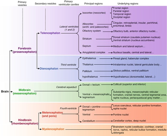
Fig. 2.4
Embryogenesis of the principal cerebral structures: from the three primary vesicles to the adult structures
The telencephalon designates an ensemble of structures that includes the cerebral hemispheres, which are subdivided into the cerebral cortex, the white matter, and the subcortical and associated structures. In phylogenetic terms, it is the most recent neural structure responsible for complex cognitive functions and is highly developed in mammals. The telencephalon is comprised of structures that are particularly implicated in olfaction, learning and memory, vigilance and attention, reward and reinforcement, pleasure and aversion, motivation, and higher affective functions.
The diencephalon underlies the two cerebral hemispheres and constitutes the structural center of the brain. It includes the thalamus, which is implicated in control of hunger and thirst, motivation, and sexual behavior. The dorsal thalamus controls the redistribution of sensory information, whereas the ventral thalamus has more of a motor role. The diencephalon is a center of integration and relay and of control of numerous functions essential for survival of the animal. It also includes the hypothalamus, which is composed of numerous small nuclei each possessing its own specific connections and neurochemical makeup. The hypothalamus is a higher center of integration of the autonomic and endocrine systems through the control that it exerts on the anterior pituitary. It is involved in major biological functions such as sleeping and waking, hunger and thirst, hormone secretion, and homeostatic functions such as blood pressure, temperature regulation, and fluid homeostasis. The epithalamus (pineal gland) regulates biological rhythms and skin pigmentation through the production and release of melatonin.
The mesencephalon, or midbrain, is a part of the brain stem composed of axon fiber bundles that are structurally and functionally connected to the rest of the brain via the cerebral peduncles, located between the pons and the diencephalon. Situated behind the peduncles are the tegmentum (the “floor” of the midbrain) and the colliculi, which are essential for visual and auditory information processing.
The metencephalon is the center for coordination of muscular movements. It is composed of the neocortical cerebellum, in the form of two cerebellar hemispheres. The cerebellum is connected to the brain stem by three pairs of peduncles: the anterior peduncle, the posterior cerebellar peduncle, and the middle cerebellar peduncle. The latter, often referred to as the pons, is a complex structure. It plays a role in locomotor function by its position as a relay between the cerebellum and the rest of the brain and participates in autonomic system function and facial sensation. It is linked to the cerebellum by the middle cerebellar peduncle and makes up the anterior surface of the fourth ventricle. The cerebellar hemispheres are linked to the neocortex by nerve pathways that control complex voluntary movements.
Related posts:


Stay updated, free articles. Join our Telegram channel

Full access? Get Clinical Tree


