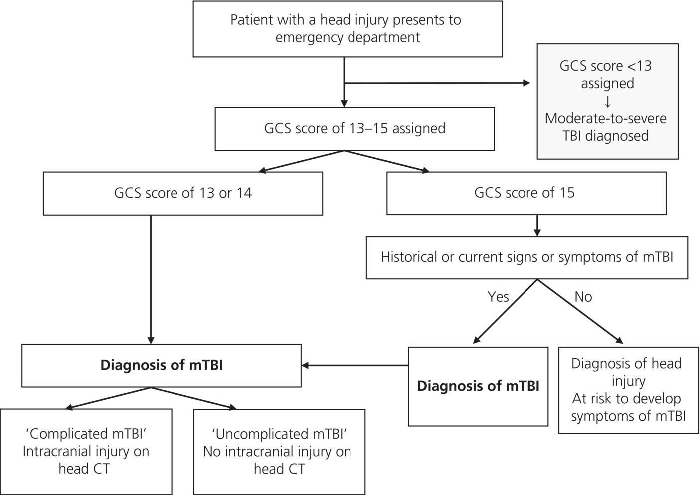
Chapter 4 Noel S. Zuckerbraun1, C. Christopher King2, and Rachel P. Berger3 1 Division of Pediatric Emergency Medicine, Department of Pediatrics, Children’s Hospital of Pittsburgh of UPMC, University of Pittsburgh School of Medicine, Pittsburgh, PA, USA 2 Department of Emergency Medicine, Albany Medical Center, Albany, NY, USA 3 Division of Child Advocacy, Department of Pediatrics, Children’s Hospital of Pittsburgh of UPMC, University of Pittsburgh School of Medicine, Pittsburgh, PA, USA Millions of people sustain traumatic brain injuries each year and for those who seek care, the Emergency Department (ED) is the front line. There are over 1 million ED visits annually in the USA for traumatic brain injury (TBI), 70–90% are mild (mTBI). Almost all of these patients will be treated and released from an ED without an in-patient admission [1]. Although mTBI may be “mild” in comparison to more severe injuries, it can still result in cognitive, physical, psychological, and social dysfunction, which can interfere with school, work, family, and social relationships, and sport participation [2–4]. Early recognition of mTBI, appropriate ED evaluation and detailed follow-up plans may facilitate recovery and reduce the risk of long-term symptoms and complications [5]. The marked variability in the definitions of and terminology for mTBI has contributed to limitations in understanding and management [6]. It is important to understand the different definitions and terminology to formulate a diagnosis and treatment plan and to communicate these findings to other medical professionals and the patient. While numerous definitions of mTBI exist in the literature, three of the most widely recognized and cited definitions are from the American Congress of Rehabilitation Medicine (ACRM), the Centers for Disease Control (CDC), and the World Health Organization (WHO) [6–8] (Table 4.1). The International Conference on Concussion in Sport proceedings offers a separate, but similar definition for sport-related concussion [9]. In all definitions, loss of consciousness (LOC) is not necessary for a head injury to be diagnosed. Indeed, 80–90% of mTBI does not involve LOC [10] and a presence of LOC does not correlate with injury severity or outcome [11]. The primary difference in the ACRM, CDC, and WHO definitions relates to the subtleties of patient inclusion criteria on both ends of the mTBI spectrum. For the ED clinician, using a less restrictive definition makes sense. This is highlighted even more in children in whom returning to sports before a complete recovery may worsen symptoms and increase the risk of reinjury [12, 13]. Table 4.1 Comparison of common mTBI definitions. mTBI is defined by trauma to the head and one or more of the following features: (i) loss of consciousness (LOC), (ii) amnesia, (iii) mental status changes as specified in the following, and (iv) other neurologic features as specified in the following. ACRM, American Congress on Rehabilitation Medicine; CDC, Centers for Disease Control; WHO, World Health Organization. * WHO definition also states that manifestations of mTBI must not be due to drugs, alcohol, and medications; caused by other injuries or treatment for other injuries (e.g., systemic injuries, intubation); or caused by other problems (e.g., psychological trauma, language barrier). While the term mTBI is being used in the current chapter, various other terms are sometimes used, including a concussion, ding or bell ringer, head injury, head trauma, closed head injury, closed head trauma, minor head injury, and minor head trauma [14–16], leading to difficulty in comparing published studies. Furthermore, when speaking with patients and families, use of some of these terms may not clearly convey the brain injury. While the CDC supports the interchangeable use of the terms concussion and mTBI [17], a recent study found that when the term concussion was used, some families interpreted this to mean that there had been no brain injury [18]. Since premature return to sports is perhaps the highest risk for children with mTBI, this misunderstanding suggests that a minor terminology change may have significant clinical significance. If ED physicians use the term concussion, it is important to stress that there has been brain trauma. Unlike more severe TBI, which typically results in significant structural brain abnormalities, mTBI is assumed to primarily cause neurometabolic dysfunction. Studies suggest that mTBI is a complex cascade of neurometabolic changes which includes an abrupt, indiscriminate release of neurotransmitters, unchecked ionic shifts, impaired axonal function, and an alteration in glucose metabolism. This cascade can result in a metabolic “energy crisis” which can persist for weeks in rat models and is believed to result in many of the clinical symptoms of mTBI [19]. However, recent studies with advanced neuroimaging (usually with MRI) indicate that subtle structural abnormalities may result from mTBI in at least some cases, particularly in athletes who are exposed to multiple such injuries [20]. The significance of these findings remains to be established. In adults less than 55 years of age, the primary causes of mTBI are motor vehicle collisions (MVC) [21]. In adults, 55 years of age and older, falls are the leading cause of mTBI. The incidence of mTBI in adults is greatest among males under 24 years of age and among men and women 65 years of age and older [22]. The leading mechanisms of mTBI in children less than 14 years of age are falls, MVC, and recreation/sports [21]. Sports-related mTBI occurs in 1.6–3.8 million adults and children annually in the USA [23]. Estimating the precise rate of sports-related mTBI is difficult due a variety of factors including underreporting by athletes and underrecognition by athletes, trainers, and clinicians [24]. Increasing access to recreational and organized sports coupled with better awareness and education in the medical, scholastic, and sport settings, will likely result in more injured athletes presenting for care in the ED [15]. Abusive head trauma (AHT)—sometimes referred to as shaken baby syndrome—accounts for over 1000 cases of TBI annually in the USA [25]. Although AHT is more frequently moderate or severe rather than mild, this is likely, in large part, due to poor recognition of AHT in its more mild forms [26]. Recognition of mild AHT in the ED can be difficult even for the experienced clinician. Misdiagnosis of AHT, even its mild forms, can have devastating consequences; it allows children to be returned to unsafe households where they can be reabused. Many of the children who ultimately die from AHT initially presented to an ED with mTBI [26]. Not every patient seen in the ED with head injury will be diagnosed with TBI. Many patients present with a focal injury (e.g., scalp abrasion), but without neurologic impairment. All patients evaluated in the ED for a head injury or injury to the body with possible force transmitted to the head should be assessed for mTBI. Common conditions, which should be trigger cues to the physician to assess for mTBI, include high-speed activities (e.g., MVC or all-terrain vehicle crash), sports and recreation, falls from significant heights, assaults, and mechanisms with external face/head injuries [17]. In addition to the patient with a chief complaint of trauma, any patient presenting with typical mTBI symptoms (Box 4.1) should have an evaluation for the presence of recent trauma. Diagnosing delayed presentations of mTBI can be challenging, as symptoms are nonspecific (Box 4.1), and the onset may occur days or weeks after injury. The initial approach to all patients with known or suspected head injury should be the same. In keeping with the American College of Surgeons Guidelines for Advanced Trauma Life Support (ATLS), the primary trauma survey should be performed with a focused assessment and stabilization of the ABCs, including cervical spine immobilization, as needed (see also Chapter 3) [27]. The Glasgow Coma Scale (GCS) is the most widely used scale to distinguish mild from moderate/severe TBI [28]. Use of the GCS in young children is challenging since the verbal score is difficult to assess. For this reason, there is a modified pediatric GCS for use in young children [29]. For the ED physician, the GCS score is most useful for the initial distinction of mTBI from moderate to severe TBI and for tracking neurologic status over time in the ED. Beyond this, its use in mTBI is limited [30]. Most ED patients with a head injury have a GCS score of 15. The role of the ED physician is to provide the necessary additional evaluation to determine which patients with head injury and a GCS score of 15 have mTBI. The algorithm in Figure 4.1 is designed to assist the ED physician in making this assessment. As discussed in the “Neuroimaging” section, not all patients who present to the ED with an mTBI will require a head CT, but those who do undergo CT can be classified as complicated or -uncomplicated based on the CT results [31]. Figure 4.1 ED process for mTBI diagnosis: from initial GCS to diagnosis of mTBI. A thorough history includes a detailed account of mechanism of injury, time of injury, immediate injury characteristics (Box 4.2), and subsequent symptoms [17]. Immediate injury characteristics are seen immediately or early after the injury and may be transient. Thus, specific inquiry to the patient, witnesses, and emergency medical services personnel as to their presence prior to ED arrival is needed. Symptoms of mTBI can be separated into four categories (Box 4.1) [17]. Headache is the most common single symptom [31]. Symptom duration is highly variable, lasting from a few minutes to several months or longer. It is also important to collect information about features of the patient’s history or injury that could change the way in which the symptoms manifest, change the risk level for an intracranial injury (ICI), and/or predict the potential for a prolonged recovery. For example, a history that medication was administered after the injury that might affect the central nervous system (e.g., opioids) and/or a history of drug and alcohol use may lower the clinician’s threshold for obtaining a CT scan. Risk factors for ICI including bleeding diathesis or known intracranial pathology should also be elicited. Finally, risk factors for prolonged recovery from mTBI should be assessed and include a history of concussion/mTBI, migraines, developmental disorders (e.g., attention deficit hyperactivity disorders) and/or psychiatric disorders [32–38]. The decision about whether to obtain a head CT in patients with mTBI is, in large part, reliant on a careful physical exam. Particular attention should be directed at the head, eyes, ears, neurologic, and skin exams. Larger and nonfrontal scalp hematomas in young children are associated with skull fracture and ICI [39]. Scalp hematomas can be boggy and subtle, especially in children with thick or braided hair. Signs of a basilar skull fracture include retroauricular bruising, periorbital bruising, and CSF otorrhea or rhinorrhea. In infants, the anterior fontanel, if open, should be evaluated for bulging, a sign of increased intracranial pressure. A detailed neurologic examination should be performed to assess cranial nerve function, motor and sensory function, and deep tendon reflexes. All injuries should be documented, particularly in cases of suspected AHT since the ED physician may be the only person to visualize concerning bruises or abrasions which can quickly fade [40]. Any bruising in a premobile infant [41] or bruising of the trunk, ears, or neck in a young child [40] should prompt concern for AHT. One of the most important clinical challenges for the ED physician is deciding whether neuroimaging is needed. A noncontrast CT is the most commonly performed neuroimaging test. Drawbacks of CT include the potential need for transporting patients outside the ED, cost, and exposure to ionizing radiation. Ionizing radiation is particularly concerning in pediatrics [42]. For the majority of patients with mTBI, neuroimaging will be normal and will contribute little to clinical management other than excluding ICI [17]. The goal is to perform the minimum number of head CTs while assuring that patients with potentially dangerous ICI are identified. For adults, numerous studies have addressed clinical criteria that physicians can use to determine whether a patient with mTBI should undergo head CT. The two most commonly used clinical criteria are the New Orleans Criteria [43] and the Canadian CT Head Rule [44]. Follow-up studies have evaluated the effectiveness of these and other criteria in terms of sensitivity and specificity for identifying clinically significant ICI in adults [45, 46]. Based on the data available from these studies, the American College of Emergency Physicians (ACEP) and the CDC issued a clinical policy statement in 2008 regarding indications for obtaining head CT scans in adults with head trauma (Box 4.3) [47].
Emergency department evaluation of mild traumatic brain injury
Defining mTBI
Definition
LOC (<30 min)
Amnesia (<24 h)
Mental status changes
Other neurologic features
GCS
ACRM [8]
Yes
Yes
Any alteration in mental state
Focal neurologic deficits, transient or nontransient
GCS 13–15, >30 min postinjury
CDC [6]
Yes
Yes
Any transient confusion, disorientation, or impaired consciousness
Observed signs of neurological or neuropsychological dysfunction, such as seizures acutely following injury among very young children: irritability, lethargy, or vomiting following head injury and symptoms among older children and adults such as headache, dizziness, irritability, fatigue, or poor concentration, when identified soon after injury, can be used to support the diagnosis of mTBI
Not specified
WHO [7]*
Yes
Yes
Confusion or disorientation
Transient neurological abnormalities (focal signs, seizure, and intracranial lesion not requiring surgery)
GCS 13–15, >30 min postinjury
Terminology
Neuropathophysiology of mTBI
Epidemiology of mTBI
ED diagnosis and management of mTBI
Use of the Glasgow Coma Scale score in mTBI

Assessment of signs and symptoms
Physical examination
Neuroimaging
Stay updated, free articles. Join our Telegram channel

Full access? Get Clinical Tree


