, Hiroyuki Tsuchiya1, Norio Kawahara1, Masahiko Hata1 and Hideki Murakami1
(1)
Kanazawa University, Kanazawa, Japan
Sacral tumors may present a difficult problem to the surgeon who desires to obtain a clear margin of excision. Frequently, tumors in this anatomical location are of a low grade biologically, such as chordomas or chondrosarcomas, and therefore unlikely to result in metastatic disease even though they are locally aggressive. Curative ablation of sacral tumors may be considered difficult because of the relationship between the anatomical location of the sacrum and the plexus of the lumbosacral nerves and vessels on the one hand and intrapelvic organs on the other. It is also difficult to reconstruct the continuity between pelvis and spine. However, en bloc sacrectomy may well be oncologically indicated, even for sacral tumors, to reduce the incidence of local tumor recurrence leading to fatal disease. In this chapter, we introduce the surgical classification of sacral tumors and the method of total or partial (segmental) en bloc sacrectomy with a T-saw [1 , 2].
Surgical Staging of Sacral Tumors
Although extensive wide excision is generally desirable for malignant sacral tumors, inadequate tumor excision is sometimes performed to prevent bladder–bowel dysfunction and loss of spinopelvic stability after sacrectomy. Because insufficient tumor ablation may cause local tumor recurrence, jeopardizing the patient’s life, complete excision of sacral tumors should be performed even though stability and the lumbosacral nerves are sacrificed.
The surgical staging system for musculoskeletal tumors was developed by Ennekinq [3] Stage 3 benign aggressive tumors, such as giant cell tumors of bone, can be treated by intracapsular excision or marginal excision. Low-grade malignant tumors, such as chordomas and chondrosarcomas, should ideally be treated with wide excision; however, at the very least, marginal excision should be performed, because an intralesional excision of those tumors will surely result in local recurrence leading to treatment failure or fatal disease [4]. High-grade tumors, such as osteosarcomas, malignant fibrous histiocytomas, and Ewing’s sarcomas, should be excised with a more radical margin beyond the reactive zone, if feasible, and treatment should include chemotherapy and/or radiotherapy.
Dysfunction After Sacrectomy
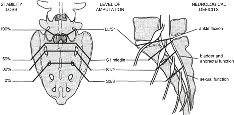
Depending on the level of sacral resection, various degrees of neurologic dysfunction and loss of stability of the sacroiliac joint will occur after the excision of sacral tumors (Fig. 37.1).


Fig. 37.1
Level of sacrectomy and dysfunction
1.
Neurologic Deficits
Sacral amputation between S2 and S3 causes sexual dysfunction because the sacral nerves below S3 are sacrificed, and sacral amputation between S1 and S2 causes bladder–bowel dysfunction because the sacral nerves below S2 are sacrificed. Total sacrectomy with the dissection between L5 and S1 leads to total loss of sacral nerve function, although ambulation can be preserved when the lumbar nerves are preserved above L5. Division of the sacrum just below the lower border of the S3 vertebra does not result in disturbance of the sphincteric function, but bilateral sacrifice of the S2, S3, and S4 nerve roots leads to urinary and fecal incontinence, and impotence for males. Patients about to undergo high sacral amputation should be apprised of this risk. Preservation of only one S2 root leads to a weakened but present sphincter control [5 , 6] whereas unilateral preservation of the S2 and S3 roots apparently has no such effect [7 , 8]. However, it is possible for patients to control urination by self-catheterization or by increasing abdominal pressure and evacuation by using an enema. The exact nature of these deficits remains controversial, however, because some authors contend that unilateral preservation of the S2 root can maintain complete anorectal continence [9] whereas others hold that when both S2 roots are preserved sphincter problems are mild and reversible [10]. Finally, it appears that early rehabilitative treatment for 1 year after surgery may restore normal bladder function [11].
2.
Stability Loss
Reconstruction should be performed depending on the degree of stability loss between pelvis and spine. Preservation of the sacroiliac joint has a great impact on the stability between spine and pelvis. In a study using cadavers by Gunterberg et al., the weakening of the posterior arch of the pelvis after sacral resection below S1 was found to be approximately 30 % and after resection through S1 approximately 50 % [12]. Native stability of the posterior arch of the pelvis is preserved, however, when sacrectomy is performed below S2. In our experience, augmentation with bone grafts and sacral rods is advisable after sacrectomy through S1 to prevent fracture of the sacral body by shearing force. After total en bloc sacrectomy, the stability between spine and pelvis should be reconstructed in combination with spinal instrumentation and bone grafts.
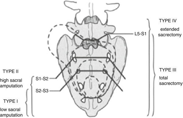
Surgical Classification of Sacral Tumors (Fig. 37.2)

Fig. 37.2
Surgical classification of procedures for sacral tumors
We classified surgical procedures for sacral tumors into four types on the basis of the extension of tumors and the level of sacral resection (sacrectomy).
1.
Type I, low sacral amputation: tumor is excised by sacrectomy below S2.
2.
Type II, high sacral amputation: tumor is excised by sacrectomy through S1 or S1–S2.
3.
Type III, total sacrectomy: tumor involving S1 is excised by sacrectomy through L5–S1.
4.
Type IV, extended sacrectomy: tumor extending beyond the sacroiliac joint or toward the lumbar spine is excised by total sacrectomy combined with excision of the ilium, vertebra, or intrapelvic organs.
Surgical Techniques
There are two major approaches for en bloc sacrectomy: the posterior approach and the combined anteroposterior approach. The posterior approach is indicated for types I and II and the anteroposterior approach for types III and IV.
1.
Type I: Low Sacral Amputation
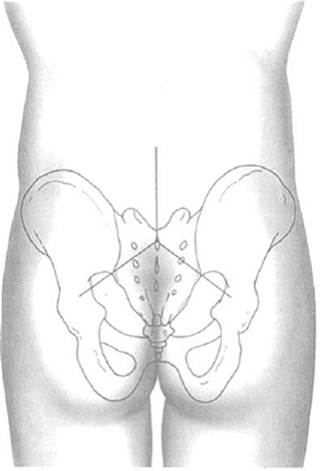
A Mercedes star incision (reversed Y) or a midline longitudinal skin incision is made on the lower back of the patient in a prone position (Fig. 37.3). The levator ani muscle and anococcygeal ligament are cut in the area around the coccyx and around the sacrum. The insertion of the bilateral gluteus maximus muscles is cut up to the edge of the sacroiliac joint. Below this, the piriformis muscle, sacrotuberous ligament, and sacrospinous ligament are cut by means of electric cauterization. The peritoneum with presacral membranous tissue is then exposed and manually separated from the presacral surface. The sacrum below the sacroiliac joints becomes free after the completion of these procedures. Laminectomy at S1, S2 is performed, and the dura mater or cauda equina is ligated and severed. A T-saw is guided from the S2 neural foramen to the greater sciatic notch, and each of the lateral wings of the sacrum is osteotomized bilaterally. Next, the T-saw is guided in front of the S2 vertebral body through the already osteotomized lines, and the S2 vertebral body is osteotomized from ventral to dorsal. For a lesion below S2, a simple transverse osteotomy with the T-saw can be conducted. In these types of cases, reconstruction is not necessary because the sacroiliac joints are not excised. However, sexual function or, rarely, bladder function is disturbed to some extent when the sacral nerves below S3 are sacrificed.


