Enlarged Perivascular Spaces
Karen L. Salzman, MD
DIFFERENTIAL DIAGNOSIS
Common
Normal Variant
Aging Brain, Normal
Less Common
Cryptococcosis
Rare but Important
Mucopolysaccharidoses
Tumor-Associated Cysts, Nonneoplastic
CADASIL
Megalencephaly with Dilated Perivascular Spaces
Hypomelanosis of Ito
ESSENTIAL INFORMATION
Key Differential Diagnosis Issues
Perivascular spaces (PVS) are pial-lined interstitial fluid-filled structures that accompany penetrating arteries
Most commonly seen as a normal variant
Helpful Clues for Common Diagnoses
Normal Variant
Round/oval fluid-filled spaces that have CSF density/intensity, no enhancement
Rare mass effect (giant PVS)
Most commonly seen at anterior commissure, inferior basal ganglia (BG)
Common: Midbrain, deep white matter (WM), subinsular cortex, extreme capsule
Rare: Thalami, dentate nuclei, corpus callosum (CC), cingulate gyrus
Aging Brain, Normal
PVS are commonly seen as the brain loses volume as part of normal aging
Helpful Clues for Less Common Diagnoses
Cryptococcosis
Enlarged PVS in BG & superior brainstem
May see DWI hyperintense rim
Helpful Clues for Rare Diagnoses
Mucopolysaccharidoses
Enzyme deficiency & inability to break down glycosaminoglycan (GAG)
PVS dilated by accumulated GAG
CC & periatrial WM most common sites
Surrounding T2 hyperintensity common ± additional patchy WM signal
Tumor-Associated Cysts, Nonneoplastic
“Cysts” caused by enlarged/obstructed PVS reported with pituitary adenomas
CADASIL
Subcortical lacunar infarcts & leukoencephalopathy in young adults
Dilated PVS are frequent in CADASIL, involving temporal WM & BG
PVS dilation in CADASIL increases with age (may be related to aging or vascular wall alterations)
Megalencephaly with Dilated Perivascular Spaces
Enlarged WM PVS with surrounding T2 hyperintensity
Hypomelanosis of Ito
Large PVS with periventricular T2 hyperintensity
Image Gallery
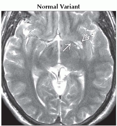 Axial T2WI MR shows a small cluster of CSF-like structures
 along the anterior commissure along the anterior commissure  at the inferior basal ganglia, the most common location for enlarged perivascular spaces. at the inferior basal ganglia, the most common location for enlarged perivascular spaces.Related posts:Stay updated, free articles. Join our Telegram channel
Full access? Get Clinical Tree
 Get Clinical Tree app for offline access

|