Fig. 15.1
Brucella colonies recovered on blood agar from blood culture of a patient with brucellosis (Courtesy of G.F. Araj, M.D.)
Brucellosis-associated central nervous system involvement is called neurobrucellosis, and while the incidence of neurological involvement changes from 3 to 25 % in various studies, it is reported to be 3–5 % in average [1, 32, 45]. Neurobrucellosis manifests with different neurological pictures as encephalitis, meningitis, myelitis, radiculoneuritis, demyelination, epidural abscess, brain abscess, spinal abscess, and granuloma [37]. Pathogenesis of central nervous system involvement is unclear, although several mechanisms have been suggested. As the bacterium Brucella lives intracellular, it avoids phagocytosis and accordingly survives for a long time. The bacteria that get rid of phagocytosis reproduce by suppression of the immunity of the host involve the central nervous system either directly or indirectly via endotoxins.
Brucellosis involves many tissues and organs, primarily the osteoarticular system, which is seen in 5–85 % of the cases [39]. The spine is the most commonly involved of bony brucellosis [26]. Brucellar spondylodiscitis begins at the upper end plates, which are rich in vascularization, depending on the virulence and inoculum of the bacteria and the immune status of the host, and may spread to the vertebral body, intervertebral disk space, and adjacent vertebra [20].
15.2 Epidural Spinal Brucellosis
Brucellar epidural abscess is encountered most frequently due to direct invasion of spondylitis, whereas it rarely develops via direct hematogeneous route without spondylitis [35]. Although brucellosis-associated spinal epidural abscesses are rarely encountered and are found in the literature usually as case reports [20, 37], their prevalence rates are reported to be 10–22 % in many studies on brucellosis [24, 26, 39, 42]. The lumbar vertebra is the most common location of involvement followed by the thoracic and the cervical vertebrae [19, 35]. Koubaa et al. [26] investigated the patients with spinal brucellar epidural abscess and demonstrated that the lumbar region is the most common location of involvement by 84.2 %.
Clinical symptoms of epidural abscess include significant spinal pain, local tenderness, and fever; however, fever may not be present in half and pain may not be present in 10–15 % of the patients [26]. Diagnosis is sometimes delayed until the appearance of neurological symptoms. Neurological complications of brucellar epidural spinal abscess are seen in 1.5 % of the cases and are usually accompanied by spondylitis [22]. Neurological complication of epidural abscess occurs due to spinal cord injury caused by direct compression effect, thrombosis, and thrombophlebitis in the adjacent veins, interruption of arterial blood flow, or the inflammation that occurs due to bacterial toxins and mediators [18]. Depending on the nerve root involved, epidural abscess leads to typical neurological symptoms such as nerve root pain defined as shooting or electric shock, motor weakness, sensorial alterations, bladder or intestinal dysfunction, and paralysis [11].
15.2.1 Diagnosis
The diagnosis of epidural spinal brucellosis is made using the combination of clinical, laboratory, and imaging methods. Brucellosis-associated epidural abscess must be considered in patients with arthralgia, high fever, perspiration, and weight loss together with epidemiological history (consumption of unpasteurized dairy products, involvement in livestock farming and veterinarian, history of past brucellosis), as well as with spinal pain, local tenderness and motor weakness, sensorial alterations, bladder and intestinal dysfunction, and paralysis. Definite diagnosis of brucellosis is made by isolating the agent from blood flow, bone marrow, or a number of other tissues. Positivity of the culture for Brucella was reported to be 41.1 % by Aygen et al. [4], 68.8 % by Colmenero et al. [9], 43 % by Kursun et al. [24], 22.6 % by Demirdağ et al. [12], 21 % by Kurtaran et al. [25], and 11.4 % by Buzgan et al. [7]. Sensitivity of blood culture ranges from 17 to 85 % depending on previous antibiotic use and duration of the disease [8, 33].
15.2.1.1 Laboratory Tests
The rose Bengal test for brucellosis is an important screening test and positive results should be verified by serum agglutination test. Quantitative standard tube agglutination test is the most common serological method used for the diagnosis of brucellosis. Detecting >160 titers in the presence of clinical findings or fourfold increase in serum antibody level in the serum samples obtained at 2–3-week intervals is diagnostic. Failing to detect seropositivity in the patients with strong clinical suspicion should suggest presence of blocking antibodies, prozone phenomenon (absence of agglutination due to excessive antibody in the patient’s serum), or early period of infection [2].
15.2.1.2 Radiological Studies
Plain X-ray, ultrasonography, computed tomography (CT), magnetic resonance imaging (MRI), and bone scintigraphy are used in the radiological diagnosis of brucellosis involving musculoskeletal system. Typical signs of brucellar spondylitis on radiography appear 3–5 weeks following the beginning of the infection [8]. The focal destructions involving the superior or inferior vertebral body angle, known as brucellar epiphysitis, are the pathognomonic signs of brucellosis. Focal anterior or diffuse disk collapse is quite common [8]. Bone erosion is less common compared to tuberculous spondylitis (also called tuberculosis of the spine). Perilesional bone formation and osteophyte formations in the anterior vertebral end plates are typical and productive osseous changes appear earlier period compared to tuberculous spondylitis. It may be difficult to radiographically distinguish it from degenerative diseases because of slow bone remodeling.
Involvement of intervertebral disk in the early period is seen as small hypodense area in the disk on CT. Flattening of disk and destruction of cartilage end plate, which cannot be detected on plain radiography in the early period, are seen on CT [31]. After administering contrast agent, paravertebral abscess and psoas involvement are easily determined. Displacement of intraspinal enlargement of epidural abscess in the posterior aspect of dural sac after contrast agent administration can be seen on CT; however, this change is identified better on MRI [30]. Sample can be obtained from disko-vertebral area by needle biopsy under the guidance of tomography. Although it is not specific, histopathological examination helps in diagnosing the brucellosis.
MRI is a scanning method used in the diagnosis and monitoring of response to medical treatment in brucellar spondylitis. In a previous study, the veteran authors of this chapter reported that MRI has high sensitivity in detecting the disease at early stage and in detecting enlargement in the epidural and paravertebral areas [39]. MRI findings in the brucellosis-associated spinal involvement show differences between acute and chronic phases. Intervertebral disk and adjacent vertebrae appear to be hypointense on T1-weighted sequences and hyperintense on T2-weighted sequences in acute brucellosis, whereas nonhomogeneous signal intensity is seen in vertebral corpus on T1-weighted images in chronic brucellosis (Fig. 15.2). Spinal involvement and paravertebral and epidural abscesses are observed as hyperintense areas due to contrast agent uptake on T1-weighted images obtained following the administration of intravenous contrast agent (gadolinium) (Fig. 15.3). Therefore, using contrast agent is critical in demonstrating spinal involvement and abscess [30, 36]. Figure 15.4 is showing spondylodiscitis (arrow) complicated with epidural abscess at the T11–T12 level.
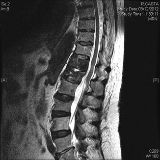
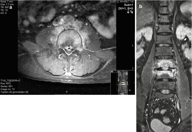
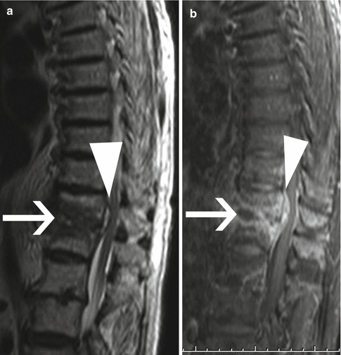
Fig. 15.2
Sagittal T2-weighted MRI reveals high signal intensity at T12 and L1, consistent with Brucella spondylodiscitis (Courtesy of M. Rodriguez, M.D.)
Fig. 15.3
MRI of a patient with brucellar spondylodiscitis. Axial postcontrast T1-weighted MRI (a) and coronal fat suppression fast spin-echo image (b) reveal the presence of brucellar spondylitis with involvement of neighboring disk space (Courtesy of Y. Ozsunar, M.D.)
Fig. 15.4
T2-weighted sagittal MRI (a) and T1-weighted fat-saturated sagittal MRI after intravenous gadolinium (b), showing Brucella spondylodiscitis (arrow) associated with epidural abscess at the T11–T12 level (arrowheads)
Bone scintigraphy is a sensitive method in detecting brucellosis-related bone involvement and is routinely recommended in the patients with musculoskeletal system symptoms [3, 15]. Bone scintigraphy allows early diagnosis of all osteoarticular involvements of the disease with a high sensitivity ranging from 69 to 91 % [3, 8]. In particular, an increased uptake limited to the anterior vertebral body angle strongly suggests presence of brucellosis [27].
15.2.2 Treatment
Eradication of microorganism forms the basis of treatment; however, there is no consensus on choice and duration of antibiotic therapy and surgical therapy. Antibiotic therapy for epidural spinal abscess is recommended for a long time ranging from 6 weeks to 1 year [20, 26, 29, 37, 40]. Antibiotics in the combinations used for the treatment of disease include tetracycline, rifampicin, aminoglycosides, trimethoprim-sulfamethoxazole (TMP/SMX or co-trimoxazole), and quinolones. Dual or triple antibiotic combinations are recommended in treatment. While neurological complications have initially been considered as indications for surgical intervention, surprisingly better responses to the medical therapy achieved in the recent times have made surgery to be considered as the last option [6]. Nevertheless, nerve root, spinal cord and dural compression, wide vertebral involvement, anterior abscess larger than 2.5 cm, and instability remain as the indications for early surgery [16, 21, 23].
Solera et al. [35] reported that they were successful with combined antibiotic therapy without surgical intervention in several cervical epidural abscess cases, which had the symptoms of spinal cord compression. Pina et al. [26] reported that they achieved cure with surgical treatment performed together with antibiotherapy due to neurological worsening in 3 of 4 patients with cervical epidural abscess. Kubaao et al. [26] investigated 19 patients with epidural abscess and reported that nine cases received dual (doxycycline and rifampicin) antibiotic combination and 10 received triple (doxycycline and rifampicin and TMP/SMX or doxycycline and rifampicin and ofloxacine) antibiotic combination for a period of 4–14 months and that none of the patients required surgical intervention and end-treatment responses were good. Faria et al. [17], Özateş et al. [28], and Ugarizza et al. [41] reported that some of the patients (36 %–89 %) required surgical treatment. Reviewing the literature, it is observed that some authors initially obtained response with medical therapy, whereas some authors obtained response with surgical intervention in addition to medical therapy. Therefore, therapy choice and duration of treatment should base on the opinions of neurosurgeons, infectious disease specialists, and radiologists depending actually on the clinical condition of the patient. The patients should be closely monitored for potential adverse drug reactions and neurological complications.
Related posts:


Stay updated, free articles. Join our Telegram channel

Full access? Get Clinical Tree


