Epilepsy

Chapter 10 Epilepsy


Epilepsy, which is a tendency to have recurrent seizures, affects about 6 of every 1000 individuals in the United States. It embodies several important neurologic problems facing psychiatrists. Seizures can mimic psychiatric disturbances and vice versa. Epilepsy is often comorbid with cognitive impairment, depression, and certain psychiatric illnesses. Moreover, many antiepileptic drugs (AEDs), which also alleviate pain, migraine, other neurologic conditions, and mood disorders, tend to induce cognitive impairment and mood changes.


Electroencephalogram (EEG)


Normal and Abnormal


Invented in the first half of the twentieth century by a psychiatrist (Hans Berger) and improved by computerization and correlation with patient videos, the EEG remains the most specific laboratory test for seizures. In addition, it still helps in the diagnosis of several other neurologic conditions. Neurologists liberally order an EEG as a painless, harmless, relatively inexpensive, and, when positive, helpful diagnostic test.


The routine EEG records cerebral electrical activity detected by “surface” or “scalp” electrodes (Fig. 10-1). Four frequency bands of cerebral activity, represented by Greek letters, emanate from the brain (Table 10-1).
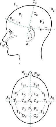


TABLE 10-1 Common Electroencephalogram Rhythms


Activity Hz (cycles/second) Usual Location
Alpha 8–13 Posterior
Beta > 13 Anterior
Theta 4–7 Generalized*
Delta < 4 Generalized*

*May be focal.


EEG readers first ascertain the patient’s age and level of consciousness. They also determine the display of the electrodes (the montage) and note the time scale, which is determined by vertical lines on the EEG paper or displayed as 1-second horizontal bars. Although approaches vary, most readers then determine the EEG’s background or dominant rhythm (see later), organization, and symmetry. EEG readers accord special attention to unusually pointed single discharges, called sharp waves or spikes, and abnormal patterns, especially if they occur in paroxysms. They judge all these features in relation to whether the patient is awake, asleep, unresponsive, or having observable seizure activity.


The normal background rhythm in an awake adult consists of waves of activity in the alpha range of 8–13 cycles per second (Hertz [Hz]) detectable mostly over the occipital region (Fig. 10-2). Neurologists refer to this pattern as the posterior dominant rhythm. It is prominent when individuals are relaxed with their eyes closed, but disappears if they open their eyes, concentrate, or are apprehensive. When people undergoing an EEG merely fix their gaze on a clock or add two single-digit numbers, faster rhythms replace alpha activity. Preoccupations, concerns, or anxiety eliminate alpha activity. Because alpha activity reflects an anxiety-free state, it represents an important parameter in “alpha training,” biofeedback, and other behavior modification techniques.
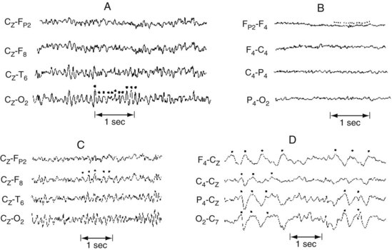


Alpha activity also disappears when people fall asleep or take psychotropic medicines. In the elderly, the background rhythm typically slows but remains within or just below the alpha range. In the early stages of Alzheimer disease, the background activity is also slower than normal. In the more advanced stages of Alzheimer disease, as well as in many other neurologic illnesses, the background EEG activity not only slows well below the alpha range, but also loses its organization.


Beta activity consists of high (> 13 Hz)-frequency, low-voltage activity located maximally overlying the frontal region. It replaces alpha activity when people concentrate, become anxious, or take many hypnotics or sedatives, including benzodiazepines. Beta activity usually inserts itself into the background EEG activity of most adults.


Theta (4–7 Hz) and delta (< 4 Hz) activities occur normally in children and everyone during deep sleep, but are usually absent in healthy alert adults. When present over the entire brain, theta or delta activity in wakefulness often indicates a neurodegenerative illness, such as Alzheimer disease, or a metabolic derangement. Continuous focal slow activity with phase reversal in bipolar montages (Fig. 10-3) sometimes originates in an underlying cerebral lesion; however, the absence of theta or delta activity certainly does not exclude one.
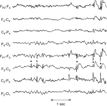


Spikes, sharp waves, and slowing – nonspecific changes – occur in about 3–15% of the general, healthy population. When they are isolated and asymptomatic, these anomalies have no clinical significance and require no further investigation. However, when spikes and sharp waves are repetitive and phase-reversed, they are an indication of an irritative cerebral focus with potential to produce seizures. Moreover, paroxysms of them suggest the diagnosis.


Seizures


During a seizure (ictus), the EEG reveals paroxysmal activity usually consisting of bursts of spikes, slow waves, or complexes of spike-and-waves or polyspike-and-waves. Although ictal EEG abnormalities are distinct when captured, muscle or movement artifacts may obscure them. After the seizure, in the postictal period, EEGs commonly show only slow low-voltage activity, postictal depression, often followed by diffuse high-voltage slowing.


In most patients, EEGs obtained between seizures, in the interictal period, contain abnormalities that support – but do not prove – a diagnosis of epilepsy. On the other hand, because many epilepsy patients have no abnormalities on an interictal EEG, a normal interictal EEG cannot exclude a diagnosis.


EEG technicians employ certain maneuvers to provoke EEG abnormalities in patients suspected of having primary generalized epilepsy (see later). For example, the technicians ask patients to hyperventilate for 3 minutes or look toward a stroboscopic light during the test. If these strategies fail to yield diagnostic EEG patterns and a strong suspicion of seizures persists, neurologists might repeat an EEG following sleep deprivation. In about 30% of epileptic patients, a sleep-deprived EEG reveals abnormalities not apparent in routine studies.


In some epilepsy patients, specially placed electrodes reveal abnormalities undetectable by ordinary scalp electrodes. For example, anterior temporal scalp, nasopharyngeal, or sphenoidal electrodes can detect discharges from the temporal lobe’s inferior-medial (mesial or medial) surface (Fig. 10-4).


image

FIGURE 10-4 Nasopharyngeal electrodes, which are inserted through the nostrils, reach the posterior pharynx. There, separated by the thin sphenoid bone, they are adjacent to the temporal lobe’s medial surface, which is the focus (origin) of about 80% of complex partial seizures. (Figures in Chapter 20 show the relatively large distance between the temporal lobe’s medial surface and the scalp, and the closeness of the temporal lobe to the sphenoid bone.) Sphenoidal electrodes are inserted through the skin to reach the lateral surface of the sphenoid wing. Electrodes in this location are near the temporal lobe’s inferior surface. (Although nasopharyngeal and sphenoidal electrodes are valuable, specially placed scalp electrodes, new arrays, electronic filters, and critical reading of the EEG may be just as accurate and less invasive.) To pinpoint a seizure focus in anticipation of its surgical removal, neurosurgeons place a grid of electrodes in the subdural space.


Another diagnostic strategy, continuous EEG-video monitoring, consists of several days of video recordings of clinical activity and EEG usually undertaken in hospital epilepsy units. The monitoring system records any seizures, changes in behavior, and effects of sleep. At the same time, physicians might check serum AED concentrations and various physiologic data. Continuous EEG-video monitoring has become the gold standard for many epilepsy studies, including diagnosing, classifying, subclassifying, and determining the frequency of seizures, evaluating patients for epilepsy surgery, treating patients who seem to suffer from refractory seizures (frequent seizures that do not respond to AEDs), and identifying disorders that mimic epilepsy, particularly psychogenic nonepileptic seizures (PNES: see later). For example, continuous EEG-video monitoring would be the ideal test for a child who has developed discrete periods of abnormal repetitive behavior in whom the routine EEG has shown only a few spikes that did not occur in paroxysms and were not temporally associated with the questionable behavior. It would also be the ideal test for an adolescent who has developed a new pattern of seizures superimposed on the usual seizure pattern in whom routine EEGs never captured a seizure.


Finally, electrodes surgically implanted in the dura, subdural space, or cerebral cortex can localize an epileptic focus when conventional tests are inconclusive. Neurologists frequently use them to pinpoint a seizure focus prior to surgery (see later).


Quantitative EEG analysis or EEG brain mapping involves topographic displays and comparisons of a patient’s EEG to standard results. This technique, still essentially a research tool, remains too unreliable for clinical usefulness in neurologic conditions, including epilepsy, minor and moderate head injury, and postconcussive syndrome. Its uncertain status precludes its credibility in litigation.


With children, an evaluation could actually begin with parents making videos of suspected seizures or other episodic disturbances, including temper tantrums, breath-holding spells, night terrors, other parasomnias (see Chapter 17), dopamine-responsive dystonia, and other intermittent abnormal movements (see Chapter 18).


Toxic-Metabolic Encephalopathy


During the initial phase of toxic-metabolic encephalopathy (delirium), when patients have only subtle behavioral or cognitive disturbances, theta and delta activity replaces alpha activity. The organization of the EEG deteriorates as the patient’s sensorium disintegrates. The EEG in toxic-metabolic encephalopathy almost always shows generalized slowing and disorganization. Additional EEG changes point to specific diagnoses. Hepatic and uremic encephalopathies characteristically produce triphasic waves (Fig. 10-5). In fact, with hepatic failure, triphasic waves often appear before bilirubin levels rise. While metabolic derangements are the most common cause of triphasic waves, this EEG finding may also be seen with toxic levels of several medications, including lithium. Benzodiazepine use produces beta activity. Herpes simplex encephalitis produces spikes and periodic lateralizing epileptiform discharges over the temporal lobes.
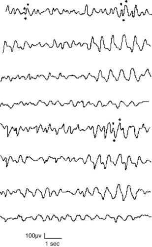


Just like a toxic-metabolic encephalopathy almost always produces EEG abnormalities, the converse holds equal weight. A normal EEG reliably precludes a toxic-metabolic encephalopathy.


Dementia


In early Alzheimer disease, the background activity usually slows to below 8 Hz. As previously mentioned, in late Alzheimer disease, the background is unequivocally slow and often disorganized.


Vascular dementia also induces EEG abnormalities. However, these changes cannot reliably differentiate vascular dementia from Alzheimer disease dementia (see Chapters 7 and 11).


In contrast, the EEG is almost definitive in diagnosing subacute sclerosing panencephalitis and common, sporadic Creutzfeldt–Jakob disease (see Chapter 7). In these conditions – characterized clinically by dementia and myoclonus – the EEG shows periodic sharp-wave complexes (Fig. 10-6). (Variant Creutzfeldt–Jakob disease [“mad cow disease”] fails to produce these EEG changes [see Chapter 7].)
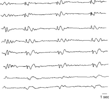


The EEG can also help distinguish between pseudodementia and dementia – to the extent that they constitute separate entities (see Chapter 7). In pseudodementia the EEG ideally would remain normal, but in dementia from almost any cause, it would show slowing. For the many patients with a mixture of depression and mild dementia, the EEG cannot measure each condition’s relative contribution to cognitive impairment.


Even though EEGs are relatively inexpensive and carry no risk, neurologists do not routinely order them in the evaluation of a patient with dementia except under certain circumstances: when dementia develops in less than 3–6 months, when myoclonus accompanies dementia (suggesting Creutzfeldt–Jakob disease or a related illness), when a patient’s level of consciousness fluctuates (indicating a toxic-metabolic encephalopathy), or if depression rather than dementia might explain poor cognitive performance.


Altered States of Awareness


The EEG shows distinctive changes during normal progressively deeper stages of sleep and during dreaming. Coupled with monitors of ocular movement and muscle activity in the polysomnogram, the EEG is critical in diagnosing sleep disturbances (see Chapter 17), which can include sleep-related behavioral disturbances and involuntary movement disorders, as well as seizures.


The EEG is also useful in diagnosing the locked-in syndrome, a condition in which patients cannot speak or move their trunk or limbs. Although patients in the locked-in syndrome appear comatose or demented, they remain fully alert and in possession of their cognitive capacity (see Chapter 11). The locked-in syndrome most commonly results from either an infarction in the base of the lower brainstem or extensive cranial and peripheral nerve damage. With their cerebral hemispheres and upper brainstem intact, patients retain normal cerebral activity and normal EEG activity.


Physicians must identify the locked-in syndrome and differentiate it from the persistent vegetative state (PVS), which is also characterized by patients’ inability to speak. PVS typically follows cerebral cortex anoxia from cardiac arrest, drug overdose, or carbon monoxide poisoning. Most importantly, because patients in a PVS have sustained extensive cerebral cortex injury, they have profound cognitive impairment. Nevertheless, their vegetative functions, such as breathing and digesting food, continue. In addition, their eyes continue to open and close, but mostly randomly or in response to light. As would be predicted, the extensive cerebral cortical damage leads to slow and disorganized EEG activity.


Finally, absence of EEG activity (electrocerebral silence), in most circumstances, indicates “brain death.” Making that determination before the heart stops beating permits procurement of organs for transplantation. Exceptions – hypothermia or drug overdose – preclude a diagnosis of brain death based on the EEG. For example, people may fully recover from either barbiturate overdose or drowning in icy water that initially left them with no obvious signs of life and a “flat” EEG.


Psychiatric Disturbances and Psychotropics


Because the EEG does not show abnormalities consistently indicative of psychiatric illnesses, obtaining EEGs on a routine basis for psychiatric patients remains unwarranted. EEGs in uncomplicated psychiatric illness show either normal patterns or frequently only minor, nonspecific abnormalities, such as excessive beta or theta activity, a few sharp waves or spikes, or poor organization.


As a confounding factor, psychotropic medications induce EEG changes. Although these changes are usually minor and nonspecific, some are prominent and persist for up to 2 months after medications are withdrawn.


There are few guidelines for assessing psychotropics’ EEG effects. Most produce background slowing into theta but sometimes delta range. Benzodiazepines and barbiturates typically produce beta activity, which may be a telltale sign of surreptitious drug use. Phencyclidine (PCP) and other excitatory drugs cause generalized, paroxysmal discharges. Phenothiazines, even at therapeutic serum concentrations, also produce sharp waves. Lithium at toxic levels, clozapine, and tricyclic antidepressants (TCAs) cause spikes and sharp waves. As described previously, lithium toxicity may induce triphasic waves on EEG monitoring. Of the antipsychotics, clozapine, olanzapine, and trifluoperazine generally produce the most EEG changes, and quetiapine, loxapine, and haloperidol, the least.


Electroconvulsive therapy (ECT) also induces EEG changes. During and immediately after ECT, EEG changes resemble those of a generalized tonic-clonic seizure and its aftermath. Subsequently, EEG slow-wave activity develops over the frontal lobes or the entire cerebrum and persists for up to 3 months. When ECT is unilateral, EEG slowing is less pronounced and more restricted to the treated side. Although ECT-induced EEG slowing is associated with memory impairment, it is also associated with more effective treatment of depression.


Seizure Varieties


The two major seizure categories are partial (or focal)-onset seizures and primary (generalized) seizures. Most partial seizures are subclassified either as partial seizures with elementary symptoms or partial seizures with complex symptoms, and most generalized seizures are subclassified as either absence seizures (absences) or tonic-clonic seizures (Box 10-1).


Partial seizures have elementary symptoms when their clinical manifestations consist of only a particular movement or sensation without alteration in consciousness. In contrast, partial seizures have complex symptoms when their clinical manifestations consist of impaired consciousness, with or without psychologic abnormalities. Both elementary and complex partial seizures originate from paroxysmal electrical discharges in a discrete region of the cerebral cortex – the focus. For example, partial seizures with motor symptoms are often attributable to a focus in the contralateral frontal cortex, and those with visual symptoms to one in the contralateral occipital cortex. Similarly, partial seizures with complex symptoms, such as psychomotor disturbances, are usually attributable to a focus in the temporal cortex but sometimes to one in the frontal cortex. Newer classification schemes eschew the descriptor “complex” because it is imprecise. Nonetheless, because psychiatrists and general neurologists routinely speak of “complex partial seizures,” this chapter will continue to use this term.


As a general rule, for the epilepsy patient, seizures consist of similar, stereotyped, individual symptoms in almost every episode. Thus, variable symptoms suggest a psychogenic disorder. Most partial seizures last between several seconds and several minutes, but in a condition known as epilepsia partialis continua or partial (focal) status epilepticus, seizures continue for 30 minutes to many hours. As long as the discharge remains confined to its focus, the original symptoms persist.


Seizures interfere with all but rote mental and physical activity, and render patients dull and immobile. Because patients can continue to perform routine activities in the midst of a partial seizure, they may continue to drive familiar routes, dress in their usual clothing, and eat.


Although seizure discharges tend to remain confined to their original cerebral cortex focus, sometimes they spread in a slow, brush-fire-like manner to adjacent cortical areas. Once the spread occurs, additional symptoms arise. Discharges may eventually spread over the entire cortex or travel directly through the corpus callosum to the contralateral cerebral hemisphere. If the discharges engulf the entire cerebral cortex (secondary generalization), patients lose consciousness, develop bilateral motor activity, and show generalized EEG abnormalities. Despite the seizure’s final, all-encompassing nature, neurologists would still classify it as a “partial seizure with secondary generalization” because they base the nomenclature on its initial manifestations.


In contrast to partial seizures, in primary generalized seizures, the thalamus or other subcortical structures seem to generate discharges that immediately spread upward, simultaneously exciting the entire cerebral cortex. Also unlike partial seizures, primary generalized seizures produce immediate unconsciousness. In the common tonic-clonic variety, bilateral, symmetric, and violent movements rile the patient, but in another common variety that occurs predominantly in children (absences, see below), no such movements occur. In any case, primary generalized seizures do not produce either focal clinical or EEG findings.


Generalized seizures, depending on the variety, usually last a few seconds to several minutes. However, both tonic-clonic seizures and absences may persist with little or no interruption for many hours, i.e., status epilepticus.


Elementary Partial Seizures


Partial seizures with elementary motor symptoms, formerly called focal motor seizures, typically consist of rhythmic jerking (clonic movement) of a body region that may be limited to one finger or extend to an entire side of the body (Fig. 10-7). These seizures may evolve into partial status epilepticus or undergo secondary generalization. Sometimes, in a “Jacksonian march,” a seizure discharge spreads along the motor cortex and creates movements, which are usually clonic, beginning in one finger and extending to the entire arm and then the face.
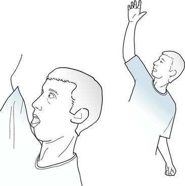


After a partial motor seizure, affected limbs may remain temporarily weak. This transient postictal paresis (Todd’s) – monoparesis or hemiparesis – may persist for up to 24 hours, though it usually only lasts minutes. Thus, the differential diagnosis of transient hemiparesis includes Todd’s hemiparesis, hemiplegic migraines, transient ischemic attacks (TIAs), and Conversion Disorder.


Seizures with elementary sensory symptoms, which usually are attributed to a focus in the parietal lobe’s sensory cortex, most frequently consist of tingling or burning paresthesias in body regions with extensive cortical representation, such as the face and thumb. Sometimes sensory loss or numbness, a “negative symptom,” might be a seizure’s only manifestation.


Elementary partial seizures with “special sensory“ symptoms consist of specific simple auditory, visual, or olfactory sensations. Although these symptoms are so vivid that they can be described as “hallucinations,” patients readily identify them as manifestations of cerebral dysfunction rather than actual events.


Patients with auditory symptoms, which are attributable to temporal lobe lesions, frequently report hearing repetitive noises, musical notes, or single meaningless words. Visual symptoms, which are attributable to occipital lesions, usually consist of bright lights. Sometimes, though, these seizures may produce lines, spots, or splotches of color that move slowly across the visual field or, like a view through a kaleidoscope, rotate around the center of vision. Physicians must distinguish elaborate visual seizure phenomena from visual hallucinations due to other causes (see Box 12-1).


Olfactory symptoms classically consist of perceiving vaguely recognizable odors, such as the frequently cited one of burning rubber. However, contrary to popular belief, these odors are not necessarily repugnant. Because olfactory hallucinations usually result from discharges in the amygdala or the uncus (the anterior inferior tip of the temporal lobe), partial seizures with olfactory symptoms are often called uncinate seizures or fits. As with other sensory symptoms, olfactory hallucinations represent the initial phase of a seizure rather than merely the warning of one. If discharges spread from sensory regions to engulf a larger area of the temporal lobe, they often trigger complex partial seizures.


EEG and Etiology


During elementary partial seizures, EEGs show spikes, sharp or slow waves, or spike-wave complexes overlying the seizure focus. For example, during seizures with motor symptoms, EEG abnormalities are usually detectable in channels overlying the frontal lobe (Fig. 10-8), and, during the interictal period, EEGs may still show occasional spikes in the same channels.
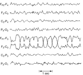


In most cases, neurologists cannot determine the cause of partial seizures. Of those cases where neurologists can establish the cause, the patient’s age at the onset of the seizures is one of the most important factors. For example, when young children develop partial seizures, typical causes are congenital cerebral malformations, such as cortical dysgenesis, and neurocutaneous disorders (see Chapter 13). In young adults, common causes of elementary partial seizures are head trauma, arteriovenous malformations (AVMs), and previously asymptomatic congenital injuries. However, posttraumatic seizures are not associated with trivial head injuries, but with serious trauma, such as trauma causing more than 30 minutes of unconsciousness, depressed (not just linear) skull fractures, intracranial hematomas, and penetrating wounds.


Young adults with major psychiatric and neuropsychiatric disorders are prone to seizures. For example, about 30% of autistic individuals and 70–90% of those with Rett syndrome develop epilepsy by the time they are adults. Also, genetic abnormalities in sodium or calcium channels or the GABAA-receptor subunit can cause both partial and generalized seizures. Such genetically determined varieties of epilepsy generally arise between infancy and adolescence. Also, in these varieties, myoclonus, mental retardation, and other stigmata of neurologic disease accompany the epilepsy.


Because drug and alcohol abuse carries multiple neurologic ramifications, neurologists often consider substance abuse in young adults presenting with seizures. Especially when seizures are accompanied by psychotic or otherwise abnormal behavior, physicians should suspect use of cocaine, PCP, and amphetamine intoxication. (Cocaine leads to seizures not only reducing the seizure threshold, but, by causing strokes, leading to noncompliance with AED regimens, and disrupting sleep.) However, although neonates may develop seizures during opiate withdrawal, adult drug addicts generally do not develop seizures during heroin use, withdrawal, or detoxification. Also, marijuana does not lead to seizures and actually has a slight antiepileptic effect.


Profound alcohol intoxication or alcohol-induced hypoglycemia can precipitate seizures. Also, 1–3 days of abstinence from chronic, excessive alcohol consumption produces seizures (see later). Although the clinical and EEG manifestations of these seizures resemble those of genetically determined seizures, the interictal EEG is normal. Withdrawal from daily use of benzodiazepines (especially alprazolam) is similar and these withdrawal seizures often evolve into status epilepticus. Most cases of benzodiazepine-withdrawal seizures are associated with prescription medicines rather than “street” drugs.


Although a seizure associated with drug or alcohol abuse may reveal dependency or addiction, it does not necessarily constitute epilepsy. Moreover, a neurologic complication of substance abuse, rather than withdrawal, may be the cause of the seizure. For example, cocaine routinely causes vasoconstriction or cerebral hemorrhages that in turn cause seizures. Similarly, although heroin may not directly produce seizures, its intravenous use can lead to bacterial endocarditis, episodes of cerebral anoxia, acquired immunodeficiency syndrome (AIDS), hepatitis, and vasculitis, which all can cause seizures.


Adults aged 40–60 years most often develop seizures because of a structural lesion, such as a primary or metastatic brain tumor. By contrast, older people are more likely to have a stroke rather than a tumor. In young adults with AIDS, the cause is most likely cerebral toxoplasmosis. The patient’s geography also suggests the cause. For example, in South and Central America, cerebral cysticercosis is the most common cause of seizures. In the Indian subcontinent, tuberculomas are one of the most common causes. By extension, those infections may very well underlie the development of epilepsy in recent immigrants to the United States.


Complex Partial Seizures


Complex partial seizures most often begin in late childhood or early adulthood. Affecting about 65% of epilepsy patients, they are the most common seizure variety. Neurologists and patients themselves solicit psychiatry consultation because of these seizures’ variable and often ambiguous ictal manifestations, interictal and postictal symptoms, and comorbid psychiatric conditions. Physicians and epilepsy patients may ask psychiatrists about antidepressants’ and antipsychotics’ indications and potential adverse reactions.


Before discussing complex partial seizures, a preliminary note on nomenclature to clarify the topic. Outdated literature applied less accurate titles, such as psychomotor seizures and temporal lobe seizures or temporal lobe epilepsy (TLE). However, neurologists apply the term “psychomotor seizures” only to the rare complex partial seizures with exclusively behavioral abnormalities. Likewise, neurologists find that the term “TLE” is not entirely appropriate because the seizure focus in about 10% of cases is located in the frontal lobe rather than the temporal lobe (see later, frontal lobe seizures). Finally, TLE is inconsistent with the current classification of seizures that is based on symptoms – not anatomic origin.


Ictal Symptoms


Symptoms of complex partial seizures in 20–80% of patients include a characteristic premonitory sensation, called an aura (Greek, breeze or soft wind). Not merely a warning, the aura constitutes the first portion of the seizure.


During most of a complex partial seizure, patients usually display a blank stare and are inattentive and uncommunicative. They always – by definition – have impaired consciousness. In most cases, they also have partial or complete memory loss, amnesia, presumably because seizure discharges besiege the limbic system in the temporal lobe. The amnesia is so striking that it may appear to be a patient’s only symptom. (Thus, physicians should strongly consider complex partial seizures among the neurologic causes of the acute amnestic syndrome [see Box 7-1].)


Physical manifestations of complex partial seizures usually consist of only simple, repetitive, purposeless movements (automatisms) of the face and hands. Present in about 80% of complex partial seizures originating in the temporal lobe, common automatisms include repetitive swallowing, mouthing, kissing, lip smacking, and lip licking – oral automatisms – or fumbling with clothing, scratching, rubbing the abdomen, or fidgeting – manual automatisms (Fig. 10-9). Other physical manifestations are simple actions, such as standing, walking, pacing, or even driving; however, sometimes these actions are simply ingrained tasks that continue despite the seizure. In addition, more than 25% of patients utter brief phrases or unintelligible sounds.
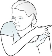


Many times the environment triggers actions and words. For example, a child may clutch and continually stroke a nearby stuffed animal while repeating an endearing phrase. Impaired consciousness, apparent self-absorption, and subsequent failure to recall the event would separate these activities from normal behavior.


Complex partial seizures occasionally cause elaborate visual or auditory hallucinations accompanied by emotions that are appropriate, inconsistent, or exaggerated. Although dramatic, such elaborate symptoms are rare.


Physicians should maintain skepticism regarding nonspecific “experiential phenomena” – déjà vu (French, previously seen or experienced), jamais vu (French, never seen or experienced), dream-like states, mind–body dissociations, and floating feelings. Having crept into the popular vocabulary, these terms have lost most of their diagnostic value. Moreover, reliable symptoms and signs and EEG findings rarely corroborate their relationship to seizures.


Another frequently encountered symptom with a dubious association with complex partial seizures is the rising epigastric sensation. This symptom consists of a sensation of swelling in the abdomen that, as if progressing upward within the chest, turns into tightness of the throat and then a sensation of suffocation. Although it could be an aura, a rising epigastric sensation may also represent a panic attack or globus hystericus, a commonly occurring psychogenic disturbance that causes a similar tightening of the throat and an inability to breathe. Likewise, when seizures originate in the amygdala, they are said to cause overwhelming fear as the primary or only symptom. However, the medical literature does not support the notion that pronounced fear, as an isolated sensation, is a seizure manifestation. More likely, it would represent a panic attack.


Many complex partial seizures, like simple partial seizures, intermittently undergo secondary generalization. In contrast, in partial status epilepticus, seizures with their same limited symptoms persist or recur in quick succession. In nonconvulsive status epilepticus, patients demonstrate hours of neuropsychological aberrations, such as thought disorder, language impairment, or change in sensorium. Patients’ behavior may be so bizarre that it merits the label ictal psychosis. It often mimics delirium, traumatic brain injury (TBI), and numerous other neurologic disorders as well as an acute psychotic episode. Neurologists usually encounter nonconvulsive status epilepticus most often in epilepsy patients, particularly in ones who have been noncompliant with their AEDs. They also see it in the critically ill patients who have sustained cerebral injury or anoxia. Neurologists diagnose it by obtaining an EEG as part of the evaluation they perform on stuporous patients. Intravenous administration of a benzodiazepine will usually abort status epilepticus and awaken the patient.


Sex, Violence, and Aggression


During seizures, patients sometimes fumble with buttons, tug at their clothing, or make rudimentary masturbatory movements. They may even seem to undress partially. However, these patients are not deliberately exposing themselves or attempting to engage in sex. Except for very rare instances, seizures are unaccompanied by erotic or interactive sexual behavior. In fact, most seizure-like symptoms that develop during sexual activity, such as lightheadedness, are simply manifestations of anxiety. (On the other hand, severe headaches that develop during sexual intercourse are ominous [see coital headache and subarachnoid hemorrhage, Chapter 9].)


Continuous EEG-video monitoring has demonstrated that ictal violence, allowing for rare exceptions, consists only of random shoving, pushing, kicking, or verbal abuse, such as screaming. This behavior is fragmented, unsustained, ineffectual, and, most importantly, unaccompanied by rage or anger. Moreover, violence, which occurs in less than 0.1% of cases, is virtually never the sole manifestation of a complex partial or any other type of seizure.


Belligerence or resistive violence, a different form of seizure-related violence, occurs when patients fight against restraints during their ictal or postictal period. Much more frequently occurring than ictal violence, resistive violence stems largely from patients’ fighting off health care workers or family members who attempt to restrain them or give them injections, as well as placing leather straps around their wrists or body.


Physicians must distinguish ictal violence, with its lack of aggression, from both criminal violence, which is characterized by aggression, and episodic dyscontrol syndrome (see later). For neurologists to consider it aggression, the behavior must be directed, have a conscious or unconscious rationale, and be accompanied by a consistent affect. Although aggression may consist only of threats or taking control, it often leads to deliberate personal and property damage.


During seizures, patients cannot engage in sequential activities, premeditated actions, or meaningful interactions with other people – requirements of criminal activity. Patients also lack the cognitive ability to operate mechanical devices. These limitations preclude violent crimes either in the midst of a seizure or as a manifestation of a seizure. Overall, most neurologists accept violence, but neither aggression nor criminal acts, as a rare manifestation of seizures.


Immediate Postictal Symptoms


Immediately after a complex partial seizure, which has an average duration of 2–3 minutes, patients characteristically experience confusion, clouding of the sensorium, disorientation, flat affect, and sleepiness. However, seizures occasionally lead not to somnolence, inactivity, and withdrawal, but to agitation, i.e., postictal agitation. If seizures involve the brain’s language region and cause transient aphasia (see Chapter 8), postictal symptoms may be more pronounced. Similarly, if the seizure focus includes the cortical areas involved with motor function, patients may have a Todd’s hemiparesis. For 15–40 minutes after a seizure, many patients have measurable physiologic changes: Approximately 40% of them have an elevated serum prolactin concentration and focal EEG depression.


Astute physicians are unlikely to mistake complex partial seizures for psychotic episodes. Complex partial seizures usually last only a few minutes, consist of stereotyped symptoms, necessarily include impaired consciousness, and usually have debilitating postictal manifestations. After recovering from a seizure and its aftermath, patients gradually return to their interictal personality, which admittedly might be abnormal. In contrast, psychotic episodes, which are frequently triggered by factors in the environment, typically last at least several days. Also, the manifestations of the psychosis vary greatly from episode to episode and often include hypervigilance.


Testing During and Between Complex Partial Seizures


EEG


During a complex partial seizure, the EEG most often shows paroxysms of spikes, slow waves, or other abnormalities in channels overlying the temporal or frontotemporal region. Even though a seizure focus may be unilateral, bilateral EEG abnormalities appear because of additional foci, interhemispheric projections, or “reflections.” Nasopharyngeal and other specially placed leads may capture temporal lobe discharges that routine scalp electrodes fail to detect (Fig. 10-10).
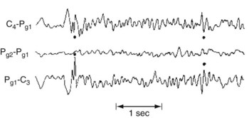


In the interictal period, the routine EEG contains spikes or spike-and-wave complexes over the temporal lobes in about 40% of cases. When accompanied by an appropriate history, these EEG abnormalities are specific enough to corroborate the diagnosis. Looking at the situation in reverse, about 90% of persons with anterior temporal spikes on the EEG will have complex partial seizures. Nevertheless, a diagnosis of seizures should not be based entirely on EEG spikes. Diagnosis of complex partial or other seizures requires correlation of EEG abnormalities with symptoms and signs.


If the diagnosis remains a problem, especially where episodic behavioral abnormalities are believed to result from seizures, physicians should arrange for continuous EEG-video monitoring. EEG corroboration of complex partial seizures might begin with a routine EEG, but it has only a 40% yield. Although EEGs performed during sleep and wakefulness or following sleep deprivation might offer a greater yield, EEG-video monitoring offers virtually a 100% yield when an event is captured.


Comorbid Conditions and Their Treatment


Depression


Depression is more prevalent in epilepsy than in other chronic neurologic illnesses, including Alzheimer and Parkinson diseases. With its prevalence in epilepsy patients ranging between approximately 7.5% and, in intractable seizure patients, 55%, depression is epilepsy’s most common psychiatric comorbidity. Given those rates, many authors assert that physicians under-diagnose and under-treat depression in epilepsy patients.


Risk factors for comorbid depression include complex partial seizures, onset of epilepsy in late adult years, and, in most studies, frequent seizures. Ironically, several AEDs – levetiracetam, tiagabine, topiramate, and vigabatrin – carry a risk for depression and self-destructive behavior (see later). In contrast, a long history of epilepsy and the laterality of the seizure focus are weak risk factors. Some studies ironically associate depression with a failure of partial seizures to undergo secondary generalization – as though experiencing a generalized seizure ameliorates underlying depression.


Once depression complicates epilepsy, seizure frequency increases. For example, depression-associated behavior, such as sleep deprivation, noncompliance with an AED regimen, or substance abuse, precipitates seizures. Depressed patients may also consciously or unconsciously superimpose PNES on epileptic ones. Additionally, depressed epilepsy patients are more likely to require hospitalization than patients suffering from depression alone.


Whatever the cause, comorbid depression worsens epilepsy patients’ quality of life. It exerts a more powerful effect than the frequency of seizures, variety of seizures, use of AEDs, or toxicity of AEDs. It is also one of several risk factors for suicide (see later).


Physicians should direct initial therapy of comorbid depression not necessarily toward depression, but toward better seizure control. Seizure control will probably improve patients’ mood, reduce behavioral disturbances, and restore some cognitive function. Fortunately, several AEDs possess mood-stabilizing as well as anticonvulsant properties: carbamazepine (which bears a structural similarity to TCAs), lamotrigine, and valproate (valproic acid/divalproex). These AEDs raise serotonin levels.


Once patients and physicians achieve optimum seizure control, they may add antidepressants to their AED regimen; however, their use carries several caveats. While antidepressants improve patients’ mood, they will probably not further reduce the frequency of seizures. Because of their effect on cytochrome P450 enzymes, psychotropic drugs may render certain AEDs ineffective on one hand or toxic on the other (see later). In the situation of depression comorbid with epilepsy, for example, prescribing an enzyme-inhibiting antidepressant, such as fluoxetine, to an AED regimen of carbamazepine or phenytoin, may lead to toxic levels of the AED. Even some apparently benign, readily available substances may alter AED serum concentrations. For example, grapefruit juice can increase concentrations of carbamazepine and zonisamide, and St. John’s wort can decrease their concentrations. Not only do AEDs potentially cause adverse drug–drug interactions, AEDs alone or in combination with other medicines may induce mental status changes (see later).


Most importantly, psychotropics, perhaps more than any other class of medication, precipitate seizures in epilepsy patients. They even cause seizures in patients with no history of epilepsy. Risk factors for psychotropic-induced seizures generally include a history of epilepsy; other neurologic disorders, including Alzheimer disease and TBI; prior ECT; and drug or alcohol abuse.


Especially with an overdose, psychotropics induce seizures. TCAs lead to seizures in approximately 5–25% of overdose cases, and the incidence following overdose of amoxapine and maprotiline is even greater. Overdose-induced seizures most often appear within 3–6 hours, but almost never after 24 hours.


Psychotropic-induced seizures, in general, most often occur during the first week of treatment, following sudden large elevations in dose, or with regimens involving multiple medicines. With routine antidepressant treatment, the risk of seizures is typically dose-dependent. For example, the incidence of seizures with bupropion immediate-release formulations at up to 400 mg daily is less than 1%, but at higher doses, the incidence rises to unacceptable levels. In an exception, clomipramine led to seizures in 1.5% of patients taking 300 mg or less per day. This relatively high rate represents clomipramine’s most significant adverse reaction. Moreover, this risk does not diminish over time, as is the case with most other antidepressants.


In contrast to the relatively high rates of seizures associated with tricyclic and heterocyclic antidepressants, monamine oxidase inhibitors, selective serotonin reuptake inhibitors (SSRIs), and other serotonin-norepinephrine reuptake inhibitors produce seizures in less than 0.3% of cases. Overall, for practical purposes, most cases of antidepressant-induced seizures result from overdose.


As a supplement or an alternative to antidepressants, ECT can alleviate depression in epilepsy patients provided that their seizures are under control. It also helps depressed patients taking AEDs; however, physicians must sometimes reduce the AED regimen before ECT. The general rule is that psychiatrists can administer ECT to patients taking an AED.


Although prolonged seizures may unexpectedly follow ECT, that complication is rare and readily responds to AEDs. (On a historical note, ECT originated in the observation that depressed epileptic patients’ mood improved after a seizure. That benefit led to physicians inducing hypoglycemic seizures by injections of large amounts of insulin. Later attempts used electricity.)


Seizures have also complicated treatment with transcranial magnetic stimulation for medication-resistant depression.


Even though most psychotropic medicines in epileptic patients are generally safe, some words of warning are required. Despite all precautions, adding any psychotropic may increase seizure frequency. Physicians can reduce the risk by slowly introducing psychotropics, attempting to use low doses of a single medicine, checking for paradoxical effects and drug–drug interactions, and monitoring serum concentrations of medicines.


On the other hand, if a patient taking a psychotropic were to develop a seizure, physicians must guard against reflexively assigning the blame to it. For example, a brain tumor might be the cause of both the seizure and symptoms of depression. Similarly, a seizure in depressed patients may result from a deliberate medicine overdose or failure to take prescribed AEDs.


Not only is depression a comorbidity of epilepsy, it is a consideration in various epilepsy-related situations. For example, seizure-like episodes are occasionally a manifestation of depression or other psychiatric conditions (see later, psychogenic nonepileptic seizures). Also, chronic depression is a risk factor for a suboptimal outcome from epilepsy surgery.


Psychosis


Besides being susceptible to postictal confusion, patients may develop a frank postictal psychosis. This thought disorder characteristically emerges after several hours to several days of clear sensorium and minimal symptoms (a “lucid interval”) following one or usually more seizures. It consists of hours to 2 weeks of hallucinations, delusions, agitation, and occasionally violence. Depending on its severity, patients usually require administration of benzodiazepines or antipsychotics.


The greatest risk factor for postictal psychosis is a preceding flurry of seizures – tonic-clonic, complex partial seizures, or both – in patients with chronic epilepsy. Up to 7% of complex partial seizures refractory to AEDs lead to postictal psychosis. Other risk factors include low intelligence, bilateral seizure foci, and a family history of psychiatric illness. Episodes of postictal psychosis, in turn, represent a risk factor for cognitive decline and interictal psychosis (see later).


Unlike postictal psychosis, interictal psychosis, loosely called “schizophreniform psychosis” or “schizophrenia-like psychosis of epilepsy” by neurologists, is a chronic condition. It generally arises when patients are 30–40 years old and their epilepsy began in childhood, especially between 5 and 10 years of age. In other words, interictal psychosis develops decades after the onset of their epilepsy. Its symptoms include persistent hallucinations, paranoia, and social isolation. Unlike typical schizophrenia patients, epilepsy patients with this interictal psychosis retain a relatively normal affect, do not deteriorate, and do not have an increased incidence of schizophrenia in their families.


Risk factors for interictal psychosis are childhood onset of epilepsy, physical neurological abnormalities, low intelligence, frequent seizures, multiple seizure types, seizures that require multiple AEDs, and episodes of postictal psychosis.


Patients with interictal psychosis also have neuropathological as well as clinical signs of brain damage. Their brains have large cerebral ventricles, periventricular gliosis, and focal damage. In an interesting comparison, multiple sclerosis (MS) patients, despite having equally extensive cerebral damage, rarely have these symptoms.


A related condition, which neurologists originally called forced normalization, followed a change in a patient’s AEDs regimen that completely suppressed abnormal EEG activity and eliminated long-standing seizures. Patients, then suddenly seizure-free, occasionally developed either psychosis or depression. Some researchers propose that the seizures, while troublesome, had suppressed a thought or mood disorder, perhaps through an ECT-like mechanism. Although the mechanisms surrounding forced normalization remain unclear, physicians should monitor patients who rapidly achieve complete seizure control. This caveat applies to epilepsy patients whose seizures suddenly come under control following epilepsy surgery or changes in their AED regimen.


In the opposite scenario, withdrawal-emergent psychopathology, after physicians or patients suddenly discontinue AEDs psychiatric disorders – particularly anxiety or depression – appear. (Although withdrawal-emergent psychopathology is a risk of suddenly stopping AEDs, status epilepticus is a much more frequent and life-threatening risk of abruptly stopping them.) Neurologists have postulated that, in patients with withdrawal-emergent psychopathology, the AEDs had suppressed a latent psychiatric disorder along with the epilepsy. Withdrawal-emergent psychopathology may also appear after epilepsy surgery – in this case because it allows patients to curtail, if not eliminate, their AED regimen.


In the preliminary version of the Diagnostic and Statistical Manual of Mental Disorders, 5th edition, (DSM-5), psychoses that occur during, immediately afterwards, or interictally would all fall into the category of Psychotic Disorder Associated with Another Medical Condition (epilepsy).


Many of the same rules apply to treating psychosis complicating epilepsy as to treating depression complicating epilepsy. Foremost, AEDs should remain the mainstay of treatment, but, if they alone are unsuccessful, neurologists or psychiatrists should add an antipsychotic. Another rule is that an overdose of an antipsychotic, just like an overdose of antidepressant, can lead to seizures. Among antipsychotics, an overdose of chlorpromazine is more likely than one of haloperidol, thioridazine, fluphenazine, or the newer atypical agents to cause seizures.


In therapeutic doses, antipsychotic-induced seizures are usually dose-dependent, occur with large increases in the dose, and develop more frequently in patients with epilepsy or underlying brain damage. In the therapeutic as well as the overdose range, chlorpromazine again remains most apt to provoke seizures. Except for clozapine, which leads to seizures in 4% of patients taking more than 600 mg daily, atypical antipsychotics carry a seizure risk of less than 1%.


Physicians forced to restart an antipsychotic following a medication-induced seizure should, while excluding other causes of psychosis and seizures, prescribe a different antipsychotic or slowly reintroduce the original one. If the patient requires clozapine or other seizure-inducing antipsychotic, physicians may offer some protection by simultaneously adding an AED.


Cognitive Impairment


Of individuals with either a congenital intellectual disability (which the preliminary version of the DSM-5 calls Intellectual Developmental Disorder, but neurologists persist in calling “mental retardation”) or cerebral palsy, 10–20% have comorbid epilepsy (see Chapter 13). In them, epilepsy usually appears before age 5 years and its incidence increases in proportion to their physical and intellectual impairments. Of individuals institutionalized because of these disorders, 40% have epilepsy. Also, children with autism and, more so, Rett syndrome are susceptible to seizures (see before).


When brain damage underlying seizures is progressive – as in tuberous sclerosis, storage diseases, mitochondrial encephalopathies, and some neurodegenerative illnesses – seizure control, cognitive capacity, and motor function all decline. Similarly, progressive cognitive decline or increasingly refractory seizures suggests a progressive rather than a congenital, static neurologic disorder. As with interictal psychosis, many risk factors for cognitive decline in epilepsy reflect underlying brain damage.


Many patients beset with complex partial seizures suffer increasingly severe cognitive impairment. Risk factors include longer duration of epilepsy, older age, and premorbid intellectual impairment. If epilepsy surgery or adjustment of AEDs controls their seizures, the cognitive decline may stop and partly reverse. However, if surgery does not arrest their seizures, their cognitive decline may accelerate. Moreover, unsuccessful surgery may lead to depression and other psychiatric disorders.


According to one explanation for the progressive cognitive impairment associated with complex partial seizures, underlying mesial temporal sclerosis leads to damage of the surrounding limbic system. Moreover, brief interictal EEG discharges in the temporal lobes probably disrupt memory and other cognitive processes.


Another, important explanation, which pertains to all seizure types and to children as well as adults, is that AEDs impair cognitive function (see later).


Crime and Interictal Violence


The consensus among neurologists is that criminal violence cannot be a manifestation of a seizure. Instead, epilepsy-related factors – intellectual deficits, poor impulse control, and lower socioeconomic status – steer people toward crime but not violence. Studies have found that, although the incidence of epilepsy is at least four times greater among men in prison than in the general population, crimes of prisoners with epilepsy are no more violent than those with PNES. Similarly, the prevalence of epilepsy is the same in nonviolent criminals as violent ones. Also, EEG abnormalities do not correlate with violent offenses.


Interictal violence rather than ictal violence, which was discussed previously, usually consists of only verbal and minor physical acts. It occurs predominantly in epilepsy patients who are antisocial, schizophrenic, or mentally retarded. Epilepsy patients with interictal violence, compared to ones without it, show no differences in the variety or frequency of seizures, EEG abnormalities, or AED treatment.


In general, behavioral changes tend to develop in individuals with a history of epilepsy that had an early onset, seizures that undergo secondary generalization, and EEGs showing bilateral changes. Epilepsy patients with comorbid psychosis or mood disorders are particularly prone to show behavioral abnormalities.


Personality Traits


Classic studies, such as those by Bear and Fedio (see References), described “temporal lobe epilepsy” patients as distinctively circumstantial in thinking, hyposexual, humorless, “sticky” in interpersonal relations, and overly concerned with general philosophic and religious questions. These patients showed excessive and compulsive writing (hypergraphia). Supporting studies suggested that the presence of these abnormal traits depended on whether the seizure focus was in the right or left temporal lobe. Right-sided foci supposedly predisposed patients to anger, sadness, and elation, but left-sided ones to ruminative and intellectual tendencies.

Only gold members can continue reading. Log In or Register to continue

Stay updated, free articles. Join our Telegram channel

Jun 4, 2016 | Posted by in NEUROLOGY | Comments Off on Epilepsy

Full access? Get Clinical Tree

Get Clinical Tree app for offline access