Evoked Potentials in Clinical Practice
QUESTIONS
1. Abnormal P100 latency can normalize in rare cases.
A. True
B. False
View Answer
1. (A): There have been some rare reports of P100 latency normalizing with or without clinical recovery. Though a rare phenomenon, it has been seen more commonly in children. (Kriss, A et al. J Neurol Neurosurg Psych 1988; 51, pp. 1253-1258)
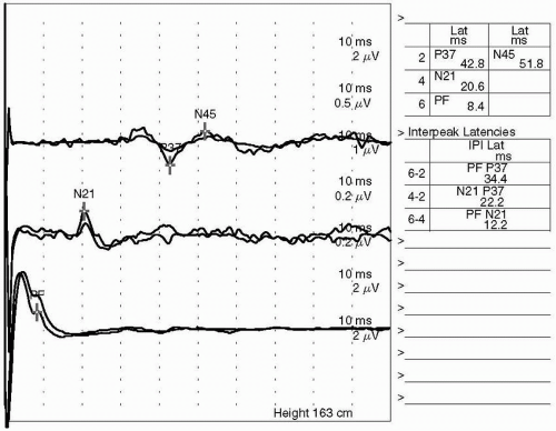
2. The findings seen in Figure 11.1 show a delay in P37 latency. This is indicative of a:
A. Conduction defect in the large-fiber sensory system above cauda equina and below the sensory cortex
B. Conduction defect in the large-fiber sensory system, peripheral to the cauda equina
C. Conduction defect in the large-fiber sensory system, peripheral to cauda equina and below the sensory cortex
D. All of the above
View Answer
2. (A): The delayed P37 absolute latency and abnormal interpeak latency of N21-P37 indicate conduction defect in the large-fiber sensory system above cauda equina and below the sensory cortex. (Chiappa 1997, p. 366)
3. The abnormal I-III interpeak latency is commonly seen with:
A. Multiple sclerosis
B. Acoustic neuroma
C. Adrenoleukodystrophy
D. Transverse myelitis
View Answer
3. (B): The abnormal I-III interpeak latency is commonly seen with acoustic neuroma, while multiple sclerosis and adrenoleukodystrophy can have abnormal III-V latency. BAEP is usually normal in transverse myelitis. (Chiappa 1997, p. 210)
4. Bilaterally delayed P100 latency can localize the lesion only anterior to the optic chiasm.
A. True
B. False
C. Bilateral delayed P100 is never seen clinically
D. Bilateral delayed P100 is pathognomonic for multiple sclerosis
View Answer
4. (B): Bilateral delayed P100 cannot be localized because it can be seen with various pathologies, for example, retinal degenerations, optic chiasm tumors, central nervous system (CNS) degenerative diseases, and bilateral optic radiation lesions. (Chiappa 1997, p. 99)
5. A 29-year-old patient presents with new onset of paraplegia. Magnetic resonance imaging (MRI) of the spine shows T2 high-intensity signal in the cord, but brain MRI is negative. Further work-up should include all of the following tests except:
A. Evoked potential
B. Echocardiogram
C. Lumbar puncture (LP)
D. Nerve biopsy
View Answer
5. (D): Nerve biopsy will not be helpful in the case of a myelopathy. However, evoked potential may help in finding other silent lesions in visual or auditory pathways suggestive of multiple sclerosis. LP will help in evaluation of inflammatory or autoimmune myelopathy, while electroencephalography (ECG) will help in ischemic myelopathy. (Chiappa 1997, p. 368)
6. Brain death is associated with which of the following brainstem auditory evoked potential (BAEP) abnormalities?
A. Absence of bilateral wave I
B. Absence of bilateral wave III
C. Absence of all waveforms following wave I bilaterally
D. All of the above
View Answer
6. (C): Without wave I present bilaterally, no inference can be made. However, in the presence of wave I, absence of BAEP waves V is compatible with brain death. (Chiappa 1997, p. 238)
7. Delayed-lumbar potential N21 but normal N21-P37 interpeak latency indicates:
A. Conduction defect in the large-fiber sensory system above cauda equina and below the sensory cortex
B. Conduction defect in the large-fiber sensory system, peripheral to the cauda equina
C. Conduction defect in the large-fiber sensory system, peripheral to cauda equina and below the sensory cortex
D. All of the above
View Answer
7. (B): Delayed-lumbar potential N21, but normal N21-P37 interpeak latency indicates conduction defect in the large-fiber sensory system, peripheral to the cauda equine. (Chiappa 1997, p. 366)
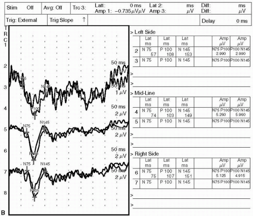
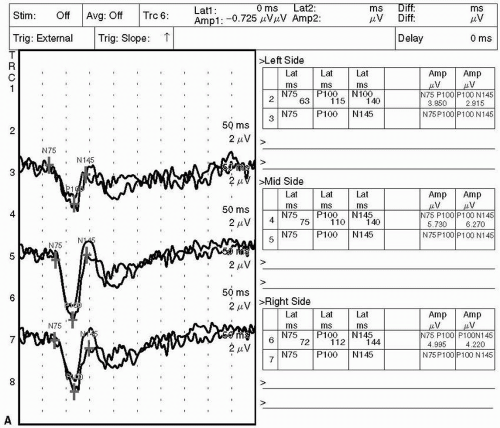
8. The tracing shown in Figure 11.2 is that of visual evoked potential (VEP) with right and left eye stimulation. These findings localize the lesion:
A. Anterior to the optic chiasm
B. Posterior to the optic chiasm
C. Cannot localize the lesion
D. Is a normal study
View Answer
8. (A): This tracing shows normal P100 absolute latency but the interside difference is significant. Unilateral P100 delay is seen with prechiasmal lesion. A unilateral P100 abnormality indicates an ipsilateral lesion of the visual pathway anterior to the optic chiasm such as a unilateral demyelinating process or optic nerve glioma. (Chiappa 1997, p. 99)
 Figure 11.2. (Continued)
Stay updated, free articles. Join our Telegram channel
Full access? Get Clinical Tree
 Get Clinical Tree app for offline access

|


