Front Neurol Neurosci. Basel, Karger, 2014, vol 33, pp 123-134 (DOI: 10.1159/000353193)
______________________
Neurosonological Examinations of Transient Ischemic Attack
Vijay K. Sharmaa· K.S. Lawrence Wongb
a Division of Neurology, National University Hospital, Singapore, Singapore; b Division of Neurology, Prince of Wales Hospital, Chinese University of Hong Kong, Hong Kong, SAR, China
______________________
Abstract
Cerebrovascular ultrasonography is the only modality that provides real-time information about blood flow in various cervicocerebral arteries. Continuous information can be obtained over extended periods with high resolution and excellent spatial display. Hemodynamic changes in the cerebral circulation due to various physiological or pathological states can be monitored reliably. The information obtained from cerebrovascular ultrasonography carries diagnostic, therapeutic as well as prognostic potential in various conditions. Cervical duplex sonography evaluates blood flow as well as arterial wall characteristics in the major arteries that supply the cerebral vascular bed. Validated criteria for the diagnosis of steno-occlusive disease of carotid or vertebral arteries have high accuracy parameters. Transcranial Doppler (TCD) ultrasonography provides a reliable evaluation of intracranial blood flow patterns in real-time and adds physiological information to the anatomical details obtained from other neuroimaging modalities. Cerebrovascular ultrasonography is relatively cheap, can be performed at bedside, and allows monitoring in acute emergency settings. Extended applications of TCD provide important information about the pathophysiology of cerebrovascular ischemia and risk stratification. Therefore, cerebrovascular ultrasonography has become an integral component of the armamentarium of stroke neurologists for understanding stroke etiopathogenesis, planning and monitoring definitive treatment and determining the prognosis. It has been suggested as an essential component of a comprehensive stroke center. We have reviewed various established applications of ultrasonography in patients with cerebrovascular ischemia.
Copyright © 2014 S. Karger AG, Basel
Transient ischemic attack (TIA) is often considered as a medical emergency that carries a substantial short-term risk of acute ischemic stroke [1, 2]. Recent advances in imaging technology have significantly improved our understanding of the etiopathophysiology of TIA. The current guidelines advocate the urgent diagnostic evaluation of all TIA patients and immediate hospitalization of high-risk cases [3].
Owing to the limited availability, risk of radiation exposure with various imaging modalities, cost and risk of nephrotoxicity with contrast agents, noninvasive imaging methods with acceptable accuracy parameters for determining the degree of arterial stenosis in cervicocerebral arteries are the preferred methods for initial screening of patients with acute cerebrovascular ischemia. Furthermore, cerebrovascular ultrasound helps in the evaluation of suspicious findings on physical examination (for example, bruits). Although vascular imaging is urgently indicated in TIA, it is not performed in time in a significant proportion of patients. Availability of diagnostic imaging methods and their cost remain the commonest limiting factors, especially in the developing world. These limitations can be overcome, at least to some extent, by using cerebrovascular ultrasonography as the primary screening modality. In addition to confirming the focal cerebral ischemia as the cause of TIA due to various steno-occlusive diseases of the cervicocerebral arterial tree, cerebrovascular ultrasonography might help in identifying suitable patients for various therapeutic interventions. While cervical duplex ultrasonography (CDU) can evaluate major arteries in the neck, transcranial Doppler (TCD) would help in understanding various hemodynamic adjustments in patients with steno-occlusive disease of cervicocerebral arteries. In this article, we would discuss the basic principles of CDU and TCD along with their advanced application in specific steno-occlusive disease states.
Cervical Duplex Ultrasonography
CDU is widely used in patients with cerebrovascular ischemia to screen the extracranial carotid and vertebral occlusive diseases [3]. CDU is relatively cheap and a bedside tool that provides important information regarding the status of vascular lumen and atherosclerotic burden in major cervical arteries. Currently available carotid imaging modalities (digital subtraction angiography, magnetic resonance angiography and computerized tomographic angiography) provide information about the arterial lumen only. CDU possesses a unique ability to evaluate the arterial wall as well as the lumen. Hence, it can be used to evaluate atherosclerotic disease burden by carotid wall imaging for intima-media thickness (IMT) and atheromatous plaques as well as for luminal imaging to estimate the degree of stenosis.
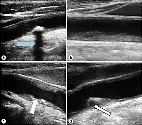
IMT is measured as the distance between lumen/intima interface to the media/adventitia interface and an increased IMT is considered the earliest sign of carotid atherosclerosis. It can be measured either manually or by inbuilt software in some ultrasound scanners. An atheromatous plaque is described as a focal increase in the IMT (more than 1.2 mm). Carotid plaques are described by their composition (echogenicity) and surface. The pivotal Tromso study showed that echolucent plaques are associated with increased risk of cerebrovascular events, independent of the degree of stenosis and other cardiovascular risk factors [4]. Hypo- or anechogenic plaques and plaques with complex pattern of echogenicity carry higher risk of cerebrovascular events than echogenic and heterogeneous plaques [5]. Various types of plaques seen on CDU are shown in figure 1. The plaque is usually covered by a thin hyperintense rim of fibrous tissue, while ‘ulceration’ corresponds to an irregularity or break in its surface. A significant ulcerations recess must be at least 2 mm deep and 2 mm long. Carotid plaque surface irregularity/ulceration exposes the thrombogenic layers, leading to thrombus formation and embolization. The guidelines for selecting the high-risk patients remain vaguely defined. Cerebral embolization from atherosclerotic plaques is determined by its ‘embolic potential’. Therefore, detection of microembolic signals (MES) on TCD may serve as a surrogate marker for stroke risk and help in identifying high-risk plaques, their embolic potential, effect of medical therapy as well as deciding the timing of various revascularization procedures.

Fig. 1. Brightness mode (B-mode) images of carotid artery and various types of plaques. a A calcified plaque produces a dense shadow (arrow) behind due to strong reflection. Tissues behind the calcified plaques cannot be evaluated due to this intense shadow. b A large smooth homogeneous plaque is noted. c Ulceration of a plaque (arrow) seen as a breach on the surface of the plaque increases the risk of local thrombus formation and distal embolization. d Sometimes the atherosclerotic plaque grows bigger, and an area can break down due to necrosis and intraplaque hemorrhage, considered to be associated with significantly higher risk of distal embolization and ischemic stroke.
Degree of carotid stenosis is the most important predictor of cerebrovascular events. CDU is often the primary diagnostic modality for carotid stenosis. CDU criteria for quantifying carotid stenosis have been validated against contrast arteriography, and for a carotid stenosis of 50-99%, it has sensitivity and specificity between 90 and 95% [6]. CDU can be used to monitor the progression of carotid stenosis in asymptomatic patients. Furthermore, it can also be used to monitor the results of carotid revascularization procedures.
Although steno-occlusive disease of carotid artery contributes most to TIA, vertebral or subclavian arteries may also be involved in some cases. CDU permits reliable evaluation of these cervical arteries too.
Transcranial Doppler
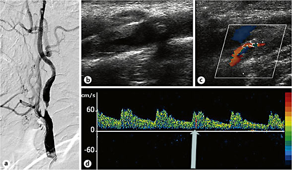
TCD can be aptly called as the stethoscope for the brain. Since its introduction in 1982, TCD has evolved as a diagnostic, monitoring and therapeutic tool in patients with cerebrovascular ischemia. Currently, TCD is the only diagnostic tool that can provide real-time information about cerebral hemodynamics and can detect embolization to cerebral vessels. Similar to CDU, TCD is noninvasive and a bedside tool and helps in obtaining information regarding the collateral flow across various branches of the circle of Willis. Compared with computed tomography angiography, TCD has 79% sensitivity and 94% specificity in detecting intracranial stenosis [7]. Advanced applications of TCD like emboli monitoring, vasomotor reactivity and detection of right-to-left shunts (RLS) help in understating the etiopathogenesis of cerebrovascular ischemia, risk stratification and prognostication (fig. 2). In the setting of acute cerebrovascular ischemia, combined TCD and CDU can evaluate the cerebral hemodynamic consequences of extracranial carotid stenosis and help in identifying lesions amenable for interventional therapy [8].
Transcranial Color-Coded Duplex
Transcranial color-coded duplex (TCCD) provides two-dimensional gray-scale real-time and color Doppler imaging of the circle of Willis. It enables the operator to obtain angle-corrected flow velocities from various intracranial arterial segments.
TCCD might be considered more accurate in the assessment of flow velocities than conventional TCD. However, no direct comparison of both methods in pathologic conditions exists to date. The flow velocities in the intracranial arteries in healthy individuals are approximately 10-30% higher when measured using TCCD as compared to conventional TCD. The difference is believed to widen in patients with in-tracranial stenosis. The comparison of the diagnostic efficiency of both methods at the same velocity thresholds cannot be valid, and it is difficult to use the flow velocities defined by conventional TCD studies for analyzing intracranial stenosis by TCCD. TCCD has been reported to have high sensitivity and specificity in the detection of a moderate intracranial stenosis. For instance, the WASID study used a mean flow velocity (MFV) threshold of 100 cm/s for middle cerebral artery (MCA) for TCD, considerably different when TCCD was used [9]. However, TCCD remains operator dependent, and the quality may be adversely affected in patients with insufficient temporal acoustic windows. Three-dimensional reconstruction during TCCD imaging and the use of sonographic contrast agents could help in improving the quality as well as reliability of the results.

Fig. 2. Imaging and neurosonology findings in a patient with recurrent TIAs. This 67-year-old male presented with recurrent episodes of left-sided weakness for about one week. Magnetic resonance imaging of the brain was unremarkable. However, the magnetic resonance angiography demonstrated a severe focal stenosis of the proximal ICA in the neck, confirmed on digital subtraction angiography (a
Stay updated, free articles. Join our Telegram channel

Full access? Get Clinical Tree


