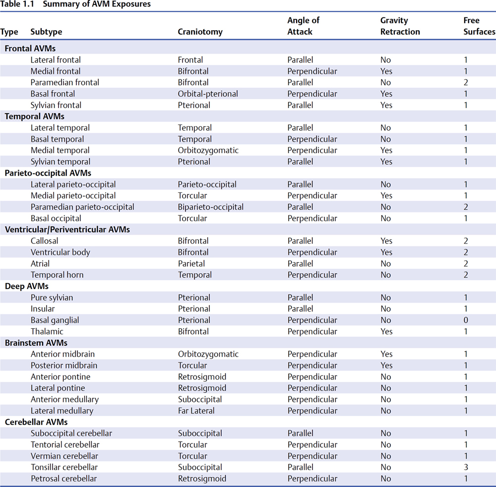
1 Arteriovenous Malformation Exposure Arteriovenous malformations (AVMs) come in many shapes: spheres, ellipses, cylinders, and, classically, cones. The cone is based on the cortical surface and tapers to a tip at the ependyma. Although AVMs are never cube shaped, it is useful to conceptualize them as a box with six sides: one top (or superficial side), four sides, and a bottom (or deep side). This orthogonal conceptualization applies structure to an otherwise amorphous tangle, giving the AVM definable sides that are medial and lateral, anterior and posterior, superior and inferior. These distinct sides can then be characterized by relationships to dural structures, cortical landmarks, feeding arteries, draining veins, fissures, and cranial anatomy. When preparing for surgery, I study computed tomography (CT) scans, magnetic resonance (MR) images, and angiograms scan by scan, sequence by sequence, and run by run, to familiarize myself with the AVM in ways that are not possible in surgery. Angiograms can replay forward and backward to see relationships between arteries and veins, to analyze bifurcations or tortuosities that may be buried in a sulcus, and to identify embolic agents that might serve as landmarks intraoperatively. Magnetic resonance images can be scrolled up and down to localize the motor strip or Broca’s area, to identify clean pial planes in a fissure, or to map out the adjacent clot as a route of access. The box enables the neurosurgeon to organize AVM anatomy as this information is gathered. A side can be “hot” or “cold” depending on its arterial input; a side can be eloquent or non-eloquent depending on its proximity to critical neurologic structures; a side can be pial, parenchymal, or ependymal, depending on its depth in the brain; and a side can be ruptured or unruptured depending on the presence or absence of an adjacent hematoma. The box concept also organizes microsurgical dissection. Hot sides are confronted early to de-arterialize the AVM, whereas eloquent sides are confronted late when the nidus is quiet. Ruptured sides are also dissected early when hematoma evacuation accesses the nidus or relaxes the brain. The box localizes specific arterial feeders. For example, paramedian parietal AVMs receive anterior cerebral artery (ACA) input on its anterior side, middle cerebral artery (MCA) input on its lateral side, and posterior cerebral artery (PCA) input on its inferior side. Similarly, the box localizes draining veins, deep tracts, and ventricles, and the box paces progress as dissection advances from top to the four sides to bottom. Most AVMs are based on a cortical surface, and this cortical surface defines the AVM subtypes. A simple example is the lateral frontal AVM based on the superior, middle, or inferior frontal gyrus. Cortical, cerebellar, and brainstem surfaces have been detailed thoroughly by Albert Rhoton and colleagues, and the names for AVM subtypes are derived from this magnificent body of work. A surface perspective generates an intuitive nomenclature that is descriptive, practical, and not overwhelming (32 subtypes). Like characterizing plants or animals by genus and species, each AVM can be characterized by type and subtype, the former based on brain location and the latter based on brain surface (Table 1.1). Arteriovenous malformation exposures access this defining surface. The lateral frontal AVM is exposed with a unilateral convexity craniotomy, and the dural opening immediately exposes the top of the box. The top side of the AVM box comes to the cortical surface and, in a sense, is already separated from the brain. “Free” surfaces require no parenchymal dissection, only the craniotomy and dural opening (free convexity surfaces, Table 1.2). They may still require the release of arachnoid adhesions or division of meningeal feeding arteries, but not the pial and parenchymal invasion required for other bound sides. Some free surfaces may not be exposed with craniotomy and dural opening alone (free fissure surfaces, Table 1.2); for example, the basal temporal AVM has a free surface inferiorly that requires subtemporal dissection for access, and the medial frontal AVM has a free surface medially that requires interhemispheric dissection for access. Most AVMs have one free surface, but some may have two or three or more, and others buried in brain parenchyma may have no free surfaces (Fig. 1.1). Table 1.2 Summary of Free Surfaces
 The Box
 Surfaces
Free Convexity Surface | AVM Subtype |
Supratentorial | |
Frontal convexity | Lateral frontal AVM |
| Paramedian frontal AVM |
Temporal convexity | Lateral temporal AVM |
Parieto-occipital convexity | Lateral parieto-occipital AVM |
| Paramedian parieto-occipital AVM |
Infratentorial | |
Cerebellar convexity | Suboccipital cerebellar AVM |
Free Convexity Surface | AVM Subtype |
Supratentorial | |
Subfrontal plane | Basal frontal AVM |
Subtemporal plane | Basal temporal AVM |
| Medial temporal AVM (posterior) |
Sylvian fissure | Frontal sylvian AVM |
| Temporal sylvian AVM |
| Pure sylvian AVM |
| Insular AVM |
Interhemispheric fissure | Medial frontal AVM |
| Paramedian frontal AVM |
| Medial parieto-occipital AVM |
| Paramedian parieto-occipital AVM |
| Callosal AVM |
| Ventricular body AVM |
| Basal ganglial AVM |
Choroidal fissure | Ventricular body AVM |
| Thalamic AVM |
Supratentorial-infraoccipital | Basal occipital AVM |
Infratentorial | |
Sylvian fissure | Anterior midbrain AVM |
Supracerebellar-infratentorial fissure | Vermian cerebellar AVM |
Tentorial cerebellar AVM | |
Posterior midbrain AVM | |
Cerebellomesencephalic fissure | Tentorial cerebellar AVM |
| Posterior midbrain AVM |
Cerebellopontine fissure | Petrosal cerebellar AVM |
| Lateral pontine AVM |
| Anterior pontine AVM |
Cerebellomedullary fissure | Tonsillar cerebellar AVM |
| Lateral medullary AVM |
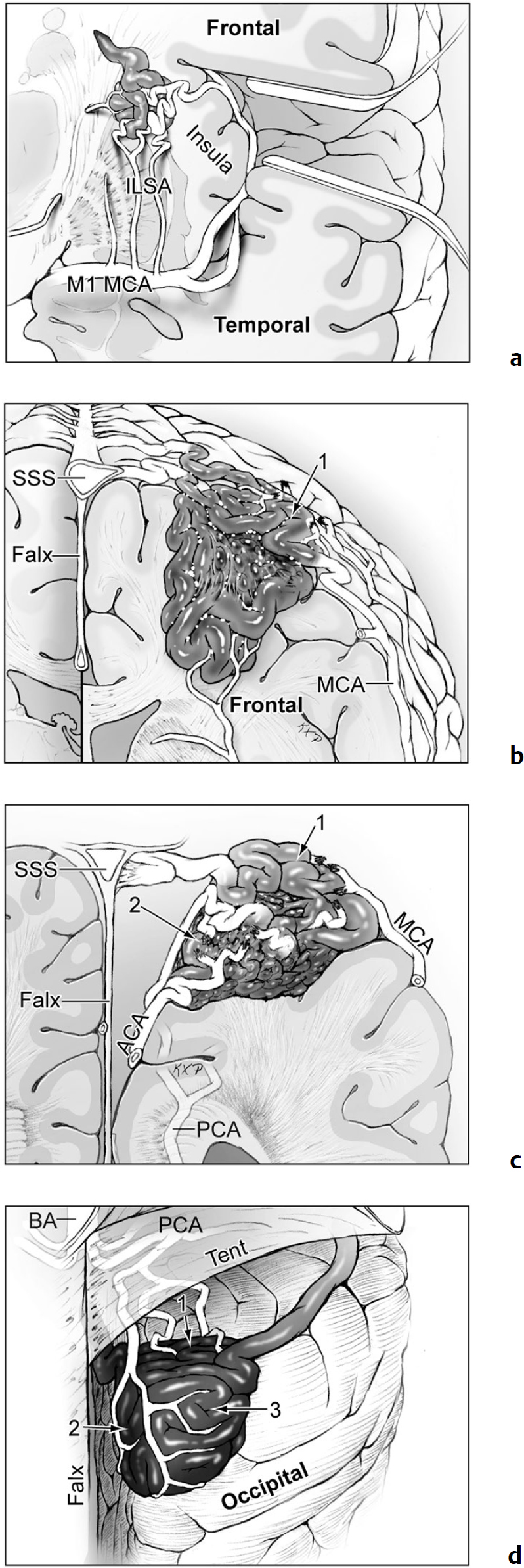
Fig. 1.1 Arteriovenous malformations (AVMs) are based on a cortical surface that defines its type and subtype, and the craniotomy exposes this surface. An AVM presenting on this surface has a free surface that requires no parenchymal dissection. Most AVMs have one free surface, but can have multiple free surfaces or none. (a) Basal ganglial AVM with no free surface (coronal cross-sectional view); (b) lateral frontal AVM with one free surface (lateral surface, coronal cross-sectional view); (c) paramedian parieto-occipital AVM with two free surfaces (lateral and medial surfaces, coronal cross-sectional view); and (d) occipital pole AVM with three free surfaces (lateral, medial, and basal surfaces, posterior view).
Stay updated, free articles. Join our Telegram channel

Full access? Get Clinical Tree


