Extra-Axial Flow Voids
James D. Eastwood, MD
DIFFERENTIAL DIAGNOSIS
Common
Normal (Normal Arteries, Veins)
CSF Pulsation
Saccular Aneurysm
Fusiform Aneurysm, ASVD
Arteriovenous Malformation
Developmental Venous Anomaly
Less Common
Dural A-V Fistula
Thrombosis, Dural Sinus
Fusiform Aneurysm, Non-ASVD
Dissecting Aneurysm
Pseudoaneurysm
Rare but Important
Vein of Galen Malformation
Venous Varix
ESSENTIAL INFORMATION
Key Differential Diagnosis Issues
Vascular vs. CSF flow void (FV)
Vascular FVs sharply demarcated from surrounding CSF
If large, vascular FVs may cause phase artifact
Vascular FV vs. acutely thrombosed artery/vein
Thrombus iso- on T1, hypointense on T2WI, can mimic FV
Do T2* (GRE/SWI) → clot “blooms”
Helpful Clues for Common Diagnoses
CSF Pulsation
Ill-defined signal loss near artery
Saccular Aneurysm
Typically involve major branch points
Fusiform Aneurysm, ASVD
Long segment & focal outpouching (BA > ICA/branches)
Arteriovenous Malformation
Extra-axial feeding, draining vessels
Look for aneurysms (feeding arteries, intranidal), venous varices
Developmental Venous Anomaly
Draining vein & enlarged medullary veins
T1 C+ best sequence
Helpful Clues for Less Common Diagnoses
Dural A-V Fistula
Older; usually prior dural sinus thrombosis
Direct AV shunt
Thrombosis, Dural Sinus
Prominent collateral veins
Fusiform Aneurysm, Non-ASVD
Often more distally located than ASVD
Vasculopathy, vasculitis, mycotic, oncotic
Dissecting Aneurysm
Extra- > intracranial; VA > supraclinoid ICA
Helpful Clues for Rare Diagnoses
Vein of Galen Malformation
↑ VOG, feeding/draining vessels
Venous Varix
Seen with AV shunting, venous strictures
Image Gallery
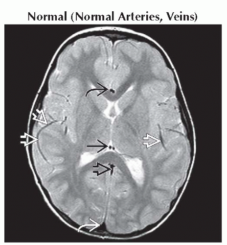 Axial T2WI MR shows paired ACA flow voids “on end”
 , curvilinear MCA flow voids , curvilinear MCA flow voids  , internal cerebral veins , internal cerebral veins  , proximal straight sinus , proximal straight sinus  , & superior sagittal sinus , & superior sagittal sinus  . .Related posts:Stay updated, free articles. Join our Telegram channel
Full access? Get Clinical Tree
 Get Clinical Tree app for offline access

|