Extradural Lesions, Multiple
Bryson Borg, MD
DIFFERENTIAL DIAGNOSIS
Common
Multiple Disc Herniations
Facet Arthropathy
Hypertrophied Ligamentum Flavum
Epidural Fluid Collections
Hematoma
Abscess
OPLL
Epidural Metastases
Plasmacytoma
Neurofibromatosis Type 1
Rare but Important
Extramedullary Hematopoiesis
Multiple Epidural Hemangioma
ESSENTIAL INFORMATION
Helpful Clues for Common Diagnoses
Multiple Disc Herniations
Disc herniations are most common ventral epidural mass in the spine
May have a thin rim of enhancement, especially if recurrent/post-operative
Facet Arthropathy
Most often associated with disc degeneration at that level
Thinning of articular cartilage, osteophyte formation
Often accompanied by ligamentous hypertrophy
“Blocky” facet morphology may be normal variant, not degenerative
Hypertrophied Ligamentum Flavum
Similar to facet arthropathy, often at a level with disc degeneration; may be a response to altered loading or instability
Posterolateral effacement of epidural fat and thecal sac
Hematoma
Post-traumatic, coagulopathic, or post-surgical etiology
Signal varies with age of hemorrhage
Mild or no peripheral enhancement
Abscess
May be associated with disc space infection or instrumentation/inoculation
Marked peripheral enhancement typical
OPLL
Thickened, calcified posterior longitudinal ligament
Ventral to thecal sac, may cause significant canal stenosis
Cervical involvement more frequent than thoracic
Best appreciated with CT
Epidural Metastases
Enhancing soft tissue mass, may be multiple
Most often due to epidural extension of a vertebral metastasis, primary epidural metastases also occur
May also occur with transforaminal spread from a paraspinal or posterior mediastinal tumor
Image Gallery
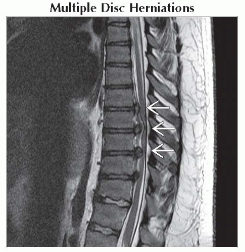 Sagittal T2WI MR shows multiple large lower thoracic disc herniations, which efface the thecal sac and compress the cord at multiple levels
 . .Related posts:Stay updated, free articles. Join our Telegram channel
Full access? Get Clinical Tree
 Get Clinical Tree app for offline access

|
