(1)
Department of Neurology, Wake Forest University School of Medicine, Winston-Salem, NC, USA
Abstract
This chapter expands fault tree analysis (FTA) introduced in Chap. 2 to medical applications. The history of FTA is discussed to introduce the reader to the diversity of fields in which it finds applications for safety and discovering the root causes of mishaps. FTA is a form of backwards thinking where the investigator starts from an event and searches for the sequence of failures which led to that event. FTA is both qualitative and quantitative, therefore it lends itself well to analysis. In this chapter, a brief review of the material introduced in Chap. 2 is presented followed by case examples where the method helped arrive at the final diagnosis.
Keywords
Fault tree analysis (FTA)Root cause analysisFailure modesFailure mechanismsRapidly progressive dementiaNeurosyphilisCADASILMyasthenia gravisAngiotrophic intravascular lymphomaGranulomatosis with Polyangiitis (Wegener’s granulomatosis)NeurosarcoidosisIntroduction
Fault tree analysis, introduced in Chap. 2 is a tool for “analyzing, visually displaying, and evaluating failure paths in a system, thereby providing a mechanism for effective system risk evaluations [1].” The history of this technique is presented here to introduce the reader to its multi-domain ubiquitous application [1]. The method was first developed by H. A. Watson of Bell Laboratories in connection with the US Air Force contract to study the Minuteman missile launch control system. Dave Haasl, then with the Boeing Company applied FTA to the entire Minuteman missile system. Subsequently, other groups within Boeing began using FTA during the design of commercial aircraft. In 1965 Boeing and University of Washington, Seattle sponsored the first system safety conference. This marked the beginning of a worldwide interest in FTA [1]. Following the lead of the aircraft industry, the nuclear power industry discovered the benefits of this technique and developed it widely. Subsequently the method was adopted by the auto industry, chemical industry, industrial automation, rail transportation, and robotics industries. The technique was used to investigate major accidents in the respective industries. The Apollo 1 launch pad fire on January 27, 1967; Three Mile Island nuclear plant accident on March 28, 1979 were investigated using FTA methods [1]. FTA tends to be used in high-risk applications as part of system safety assessment [1]. Major applications of the technique include: verifying numerical requirements, identification of safety critical components, product certification, product risk assessment, accident/incident analysis and design change evaluation, visualizing causes and their consequences, and common cause analysis [1]. The interested reader will find a wealth of information in references [2, 3] and in Chap. 2.
This chapter explores FTA for medical diagnosis which is a form of accident investigation. Starting from symptoms, physical examination, and laboratory evaluation data, we would like to work backwards to the root cause(s) of what we observe. Once we identify the cause(s), we would like to be able to reconstruct all the events which led to the incident under investigation. For the purposes of making this section self-contained, important principles of constructing a fault tree are reviewed here followed by diverse case examples.
Constructing a Fault Tree
The following events and logic gates will be used in the medical case examples discussed in this chapter [2, 3]:
A. Primary events: of a fault tree are those which are not developed further. The most important primary events for the purposes of this chapter are:
A.1: Basic event: This is a basic initiating fault which does not require further development.
Medical example: (a) Herpes Simplex is the cause of encephalitis in this patient, (b) myasthenia gravis is the cause of weakness. The investigator therefore needs to define the limits of resolution of his analysis to define the basic event. For the myasthenia gravis example above, we do not extend the analysis further to the next level to define what is the cellular mechanism of weakness from myasthenia gravis.
A.2: Undeveloped events: An event which is not developed further, either because developing it further is not relevant for the problem being analyzed or because more information is not available. This usually helps direct investigations and prevents distractions from abnormal lab data which may not be relevant for the investigation on hand. It is represented by a diamond.
Medical example: (a) Thyroid cyst identified on MRI Cervical Spine in a patient with paralysis of the lower extremities. (b) Benign renal cyst on MRI Lumbar Spine performed for foot drop. (c) A Vitamin B12 level of 400 in a patient with paraplegia.
B. Intermediate event: a fault event that happens because of one or more primary events acting through logic gates. It is represented by the rectangle symbol and represents an intermediate step in the analysis.
Medical example: (a). Amyotrophic lateral sclerosis (basic event) led to diaphragm weakness (intermediate event) which led to ventilatory failure. (b) Myasthenia Gravis (basic event) led to pharyngeal weakness (intermediate event) which led to aspiration (intermediate event) which led to pneumonia.
C. Logic Gates: are the logical combinations of primary and intermediate events (building blocks of the tree) which lead to the undesired top event. The following gates are described here since they find application in the medical case examples discussed in this chapter:
1.
Boolean “OR” gate: The output occurs if at least one of the input events occurs.
Medical examples: (a) Hypothyroidism OR Myasthenia led to Weakness. (b) Cardiac failure or COPD caused shortness of breath.
2.
Boolean “AND” gate: The output event occurs if and only if all the input events occur. Either one or the other input events cannot cause the output to happen.
Medical example: (a) Diastolic dysfunction AND fluid overload led to pulmonary edema.
3.
The Inhibit gate: represented by a hexagon is a special case of the AND gate. The output can be caused by a single input, but a qualifying condition must be present for the output to happen. The qualifying condition is represented by a type of primary event called the conditioning oval drawn to the side of the inhibit gate. This can be used to visualize the effect of drug interactions.
Medical example of Inhibit Gate: (a) Patient on stable dose of carbamazepine became toxic when fluconazole was added for fungal infection [4]. This happened because fluconazole (conditioning event) inhibits hepatic enzymes which are concerned with carbamazepine metabolism leading to toxicity.
The corresponding symbols for the events and logic gates are discussed in detail in Chap. 2. In this chapter, we will use English letters to denote logic gates for ease of discussion. The following rules will be followed for constructing a tree:
1.
State the undesired top level event in a clear, concise statement. Examples include “Patient is weak and numb below the waist.”
2.
Develop the upper and intermediate tiers of the fault tree: Determine the immediate, necessary, and sufficient causes to explain the top event and interconnect them by the appropriate logic symbols [2, 3]. For the top event example of “Patient is weak and numb,” the immediate, necessary, and sufficient causes are it could happen because of diseases of spinal cord OR diseases of nerves. This is called the “Think Small” rule. The investigator identifies only the immediate causes of the top event, not the root causes. Failure modes are identified followed by failure mechanisms as demonstrated in Chap. 2. To the extent possible, at each step failure modes (the manner in which the system has failed) are identified first, followed at the next level of the tree by failure mechanisms (the causes which can lead to the corresponding failure mode.)
3.
Extend each fault event to the next lower level. The immediate causes of the top event are the subtop events linked together by logic gates. Each subtop event becomes the top event for the next level of the tree. For each subtop event, identify the immediate, necessary, and sufficient causes for the subtop event to happen. For the example above, diseases of the spinal cord can happen due to inflammatory conditions, infectious conditions, vascular diseases of the cord, etc. At each level of tree construction, particular attention is paid to the following:
Can any single failures cause the event to happen?
Are multiple failure combinations necessary for the event to happen?
In medical fault trees, we can incorporate results of available investigations and develop certain branches of the tree and stop others (undeveloped event). For example, let us assume an EMG study of the above patient shows a severe neuropathy. In that case, disease of spinal cord becomes an undeveloped event.
4.
Develop each event through its immediate, sufficient, and necessary causes till the limit of resolution is reached and root cause(s) are established. Root cause analysis is explored further as a management method in Chap. 8 of this book. For the example above, we stop developing the spinal cord branch of the tree and explore the immediate, necessary, and sufficient causes of the severe neuropathy. This may be due to immunologic causes like Guillain-Barré Syndrome or toxic causes such as heavy metal poisoning and we design appropriate tests for the same (lumbar puncture for GBS and 24 h urine heavy metal screen for toxic neuropathy). At this stage we have identified the root cause of the top event.
5.
Evaluate the fault tree in qualitative and/or quantitative terms. Fault trees are qualitative by nature of their construction. At this stage, we can rank root cause(s) in order of probabilities. For the example above, except under unusual circumstances, the probability of Guillain-Barré Syndrome exceeds that of heavy metal poisoning. Therefore, the investigator can direct his attention and further tests to this hypothesis and consider a spinal tap looking for albuminocytologic dissociation.
6.
Once the root cause(s) are identified, the investigator must be able to reconstruct the top event by traversing up the tree. The idea behind the analysis is to identify the “minimal cut set.” A “minimal cut set is the smallest combination of component failures which if they occur will cause the top event to happen” [2]. Therefore, starting from the root cause and walking forwards, the investigator must be able to reconstruct the intermediate events and finally the top event.
Medical Case Examples of Applications of FTA
The first step to performing fault tree analysis is to express the clinical problem in a simple form which forms the starting point of this method.
Case Example 1
P.D. is a 35 y/o male who started exhibiting behavioral changes approximately 2 years ago. Adopted as a child, he was employed as an engineer till matters became increasingly difficult for him because of behavioral abnormalities. This resulted in divorce, a house fire, fights with neighbors, and subsequently loss of employment. He was treated for bipolar disorder with later development of dizziness, poor balance and falls, and oral dyskinesias, possibly a side effect of antipsychotics. Progressive behavioral changes led to nursing home confinement and increasing use of restraints.
Physical examination revealed a severely confused state. Patient had severe impairments in speech, memory, attention, and was unable to follow simple commands. Cranial Nerve examination was normal. Motor strength and gross sensation were normal. Physical examination was consistent with a severe dementia involving multiple domains of memory, executive function.
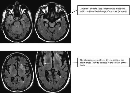
An MRI Brain done at this stage showed severe abnormalities shown in Fig. 3.1. The key features of this MRI Brain study are the severe global atrophy, a pattern of white-matter signal changes involving the bilateral anterior temporal poles, external capsule and subinsular regions. This constellation of findings raises concerns for CADASIL—cerebral autosomal dominant arteriopathy with subcortical infarcts and leukoencephalopathy [5]. No contrast enhancement was seen in this study. The referring physician had ordered the corresponding gene test involving the Notch3 gene on chromosome 19. The gene test has >95 % sensitivity and 100 % specificity [5]. Despite such a suggestive MRI Brain the gene test was negative. Therefore, one of the concerns for the referring physician was whether this patient would benefit from a skin biopsy to diagnose the condition since CADASIL causes deposition of granular osmiophilic deposits around smooth muscles of arterioles in many tissues. These deposits can be seen on electron microscopy and can be used to make the diagnosis of CADASIL when the genetic test is negative [5].


Fig. 3.1
MRI images of a 35-year-old male with rapidly progressive dementia. The pattern of signal changes involves certain key areas of the brain including anterior temporal poles, external capsule, and subinsular regions which can be seen with CADASIL [5]
CADASIL is unfortunately not treatable. Limited laboratory data available for review showed a normal result for HIV and Hepatitis B and C viruses. Since this patient’s care was fragmented across many institutions, no further data was available. A solution to this case was attempted using FTA shown in Fig. 3.2.


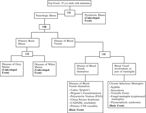
Fig. 3.2
FTA for Case Example 1. The top event is “35-year-old male with rapidly progressive dementia.” Using the “think small rule,” the investigator works his way down to the root cause(s) of this event and directs confirmatory investigations accordingly
1.
Top Event: Formulate the problem in simple terms: 35 y/o male with rapidly progressive dementia.
2.
Sub-events immediately leading to Top Event: The immediate, necessary, and sufficient conditions leading to the top event are neurological illness OR psychiatric illness. The abnormal MRI excludes a primary psychiatric illness; therefore this is an “Undeveloped event” and will not be developed further.
3.
Sub-event: Neurological Illness will be developed to the next level. The abnormalities seen on MRI Brain can be due to a (a) disease of the Brain itself OR (b) due to a disease of blood vessels. CADASIL was explored as a cause of disease of blood vessels.
4.
Sub-event: (a) Disease of the brain itself can be due to a disease of white matter OR disease of gray matter. MRI shows predominantly white-matter involvement, therefore this is developed further and diseases of grey matter (termed poliodystrophy) will be considered an “Undeveloped Event” and not developed further.
5.
Sub-event: Diseases of blood vessels can be diseases of blood vessels themselves OR involvement of the blood vessels from chronic inflammation around the surface of the brain—chronic meningitis. Available clinical information did not suggest an ongoing multisystem disease with involvement of lung, liver, or kidneys. Therefore the blood vessels covering and penetrating into the brain could be affected due to inflammation or infection around the brain from chronic meningitis.
6.
Sub-event: Primary diseases of blood vessels termed “vasculitis,” can be isolated to the brain (“primary CNS vasculitis) or be part of a multisystem autoimmune disease like SLE, Wegener’s granulomatosis, or polyarteritis nodosa [6]. Given the long duration of illness in this patient, chronic meningitis with this duration of survival without treatment can happen from diseases such as sarcoidosis, syphilis, herpes zoster, fungal (cryptococcal), and tuberculosis infections, the latter two being extremely unlikely [7].
FTA led to the bottom right of the tree since most other conditions along the way were analyzed and felt to be not relevant for the clinical presentation on hand and left as undeveloped events. The formulation has the advantage of permitting the investigator to return to any of these trains of thought and expand them should initial inquiries prove unrewarding.
Following identification of candidate root cause(s), confirmatory tests can be planned for making the final diagnosis. The candidate diagnoses are analyzed further based on available clinical information. For case example 1, this can be performed rigorously using the Bayesian approach (please see Chap. 2 for more details.)
1.
Chronic Meningitis Syndromes: We can now determine the probability of the individual diagnosis identified under chronic meningitis syndromes and rank them in descending order of probability to direct investigative resources. Starting with syphilis, we are interested in calculating the joint probability of syphilis and rapidly progressive dementia in this patient, expressed as P(Syphilis AND Rapidly Progressive Dementia). This is expressed as:
P(Syphilis, Rapidly Progressive Dementia) = P(Syphilis) × P(Rapidly Progressive Dementia ∣ Syphilis).
P(Syphilis) is a measure of the probability of syphilis. P(Rapidly Progressive Dementia ∣ Syphilis) is a measure of how likely it is to get rapidly progressive dementia from untreated syphilis.
Similarly, we can calculate the remaining probabilities:
P(Fungal Meningitis, Rapidly progressive dementia) = P(Fungal meningitis) × P(Rapidly progressive dementia ∣ Fungal Meningitis).
P(Sarcoidosis, Rapidly progressive dementia) = P(Sarcoidosis) × P(Rapidly progressive dementia ∣ Sarcoidosis).
P(Paraneoplastic Syndrome, Rapidly progressive dementia) = P(Paraneoplastic Syndrome) × P(Rapidly progressive dementia ∣ Paraneoplastic Syndrome).
2.
Diseases of Blood Vessels: In a similar manner, we can calculate the respective diagnostic probabilities of this branch of the tree.
P(SLE vasculitis, Rapidly Progressive Dementia), P(Wegener’s, Rapidly Progressive Dementia), P(Primary CNS vasculitis, Rapidly Progressive Dementia) can all be respectively analyzed in this manner. The numbers themselves can be obtained from review papers on these topics, however we are looking for a qualitative feel for the concerned probabilities.
Ranking all the probabilities, we find that P(Syphilis) × P(Rapidly Progressive Dementia ∣ Syphilis) is likely higher than the rest since syphilis is a common infection and untreated syphilis caused neurosyphilis which is a cause of severe dementia. In descending order, we get systemic vasculitis like SLE, Wegener’s and Sjogren’s syndrome. Primary CNS angiitis is much rarer; therefore it can be investigated if the above approach fails. Based on the results of the FTA, resources can be directed for the next round on testing:
The following tests were requested: Step 1: In Blood: Rapid Plasmin Reagin (RPR), Antinuclear Antibodies (ANA), Antineutrophil Cytoplasmic Antibodies (ANCA). Step 2: The next step would be testing of the CSF for infections and inflammation. Finally if these do not yield any results, a brain biopsy looking for primary CNS vasculitis can be performed. Each of these hypotheses is a valid minimal cut set. Starting from any of these basic events, we can walk back up the tree and reconstruct the intermediate events leading to the top event.
Serum RPR was positive in very high titer (>1:128). Confirmatory testing for syphilis with FTA-ABS was also positive. A directed examination of spinal fluid showed a positive CSF VDRL confirming neurosyphilis as the root cause of the patient’s rapidly progressive dementia. He was treated with high-dose IV penicillin with considerable improvement by the end of the treatment period. He no longer required antipsychotic medications or restraints. At the end of one month he was discharged home to his family with remarkable improvement. The diagnosis in this patient immediately resulted in testing of all contacts by public health authorities.
This case highlights the importance of the methodical, “one small step” at a time approach which is extremely important in constructing fault trees.
Case Example 2
J.W. is a 40 y/o male referred for myasthenia gravis. Patient reports that about 3–4 years ago he developed vertical/diagonal diplopia. This is basically constant all day long and will occasionally get somewhat better after some rest. This is worsened by horizontal gaze, primarily with right gaze. This has remained relatively stable since it began about 4 years ago, however he does state that the images have become further apart over the years. The diplopia completely resolves with the closing of either eye. He denies ptosis or extremity weakness. He does state that he feels his right eyeball is weak and he has grittiness/dryness primarily in the right eye. The patient did have a blow to the head around the time his symptoms began.
Patient first sought care from an eye doctor who felt he had extraocular muscle weakness. He then saw a neurologist in April 2013. He had a CT Chest which did not show evidence of Thymoma or other malignancy. MRI Brain without contrast was obtained and was normal. Blood tests showed: AchR Binding Antibodies: 0.26 (normal <0.25), AchR blocking antibodies negative, AchR modulating AB negative; B12, Folate, TSH, Free T4 normal; ESR 2, CRP 0.6. Patient was initially treated with Pyridostigmine but this did not provide benefit. He was then given IVIG 2 g/kg over 5 days in the last week of April. He saw no improvement with the IVIG. He was placed on prednisone which again showed no benefit. He developed significant side effects from prednisone including rash and weight gain. Since he was a professional truck driver with an international logistics company, he had been unable to work and was on disability.
On focused neurological examination, cranial nerve examination did not show ptosis. The patient had diplopia on right gaze which worsened with left head tilt with weakness referred to the left superior oblique muscle. All other extraocular muscles appeared normal at bedside clinical examination. The remainder of the neurological examination was normal. The reason for referral was for initiating plasmapheresis since IVIG, high-dose prednisone and Pyridostigmine were ineffective. A prior treatment trial with prism glasses had not been successful.
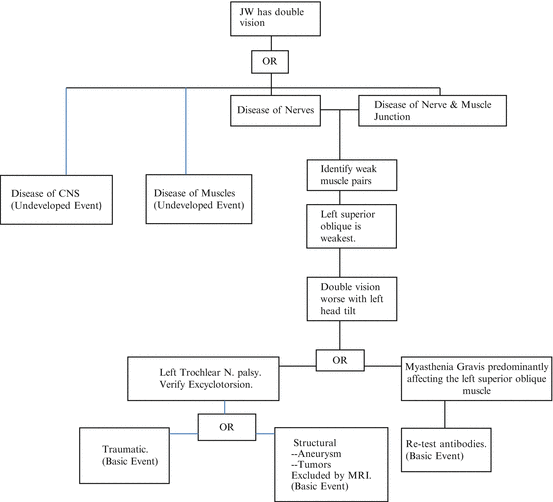
The problem was formulated using FTA methodology with the following results shown in Fig. 3.3:


Fig. 3.3
FTA for Case Example 2. By successive application of the “Think Small” rule, the two candidate root cause(s) are left trochlear nerve palsy vs. myasthenia gravis involving the left superior oblique muscle. Confirmatory testing can then be designed to discriminate between the two
Step 1: Top Event: JW has double vision.
Step 2: The immediate, sufficient, and necessary causes on the top event are: Diseases of the central nervous system OR diseases of extraocular muscles OR cranial nerves 3, 4, and 6 (especially 4 based on examination) OR neuromuscular junction disorders.
Step 3: Sub-event: Diseases of the central nervous system have been excluded by normal MRI Brain scans. Therefore this is an “undeveloped event.” Disease of extraocular muscles are genetic (example mitochondrial disorders), highly unlikely to present this way since they involve multiple muscles and also are associated with ptosis. Therefore this too is an undeveloped event. The following sub-events are chosen for expansion.
Diseases of Nerves: Involving right eye, left eye, or both.
Diseases of neuromuscular junction: myasthenia gravis, Lambert Eaton myasthenic syndrome.
Step 4: Sub-events: Disease of Nerves: Identify all weak muscles based on where images are maximally separated. This showed weakness mostly involving the left superior oblique.
Sub-events: Disease of neuromuscular junction: identify all weak muscles.
Stay updated, free articles. Join our Telegram channel

Full access? Get Clinical Tree


