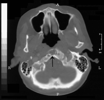
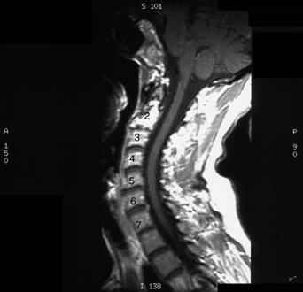
78 A 67-year-old woman complained of upper neck pain worsened by neck extension, radiating into her left sub-occipital region. Objectively, she has no focal neurologic deficit and a mild decrease in cervical range of motion. Computed tomography (CT) and magnetic resonance imaging (MRI) of the cervical spine demonstrated a destructive bony lesion involving the inferior part of C2, C1, and the clivus (Figs. 78-1 and 78-2). FIGURE 78-1 CT showing destructive bony lesion of C2. Skeletal dysplasia The patient was presented with four treatment options: (1) observation with medication, (2) biopsy, (3) posterior upper cervical fusion, and (4) vertebroplasty. She has similar lesions in her right arm and pelvis.
Fibrous Dysplasia
Presentation
Radiologic Findings

Diagnosis
Treatment

Fibrous Dysplasia
Only gold members can continue reading. Log In or Register to continue

Full access? Get Clinical Tree


