♦ Preoperative
Operative Planning
- Magnetic resonance imaging: review images with and without contrast. For operative planning, obtain postcontrast images in all planes. Carefully inspect for vessel encasement, particularly the vertebral arteries, degree of brain stem mass effect and/or edema, hydrocephalus, and syrinx of the spinal cord.
- Computed tomography: helpful to assess for any tumor calcification and/or bony changes, such as erosion of the occipital condyle or lateral mass of the atlas
- Angiogram: may aid in complex cases; potential embolization for large tumors
- Balloon test occlusion may be helpful to assess tolerance of potential loss of vertebral artery
- Exposure planned according to superior and inferior extent of tumor
- Lower cranial nerve function at baseline should be assessed preoperatively
- Steroids for significant edema
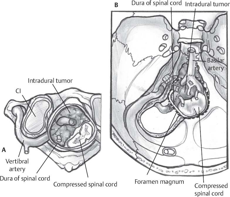
- Majority of these meningiomas are located in the anterior or anterolateral regions of the foramen magnum, requiring a far lateral approach. A posterior midline low suboccipital craniectomy with C1 laminectomy may be used for posterior and posterolateral lesions.
Equipment
- Mayfield head holder
- High-speed drill
- Ultrasonic aspirator
- Microscope
- Somatosensory evoked potentials, lower cranial nerve neurophysiologic monitoring
- Doppler: to define the course of the vertebral artery
Anesthetic Issues
- Anesthesiologist must be made aware to use agents with minimal paralysis for neurophysiologic monitoring
- Arterial and central venous lines
- Intravenous antibiotics and steroids
- Mannitol (0.5 to 1.0 g/kg) for large tumors
♦ Intraoperative
Positioning and Surgery
Far Lateral Approach (Fig. 39.1)
- Head is positioned so the mastoid ipsilateral to the lesion is at the highest point (side chosen based on tumor lateralization)
- Avoid excessive head turning to prevent venous kinking and vertebral artery stretching; be careful about stretching the brachial plexus
- Incision (hockey stick) starts at tip of ipsilateral mastoid and continues above the superior nuchal line and curves to the midline down to the level of C3. It is helpful to preserve a muscle cuff at the level of the superior nuchal line for closure and prevention of a cerebrospinal fluid (CSF) leak. May use the Doppler to localize the vertebral artery during dissection.
- Bone flap is created according to surgeon’s preference
- Dural opening is either cruciate or T-shaped (tailored to the tumor)
Only gold members can continue reading. Log In or Register to continue


