Frontal Lobe Epilepsy


Frontal Lobe Epilepsy


Introduction

The frontal lobe is the most frequent site of focal epilepsy within the extra-temporal regions. Based on this definition the onset of epilepsy is focal and not generalized and surgical resection of an epileptogenic zone in the frontal lobe would result in a surgical cure.

In patients who have medically intractable epilepsy who present to epilepsy surgical centres, approximately 10-20% of them can be expected to have frontal lobe epilepsy; however, the actual prevalence of frontal lobe epilepsy in the general population may be significantly larger. The actual prevalence is difficult to ascertain due to the difficulty in localizing the epileptogenic zone in many of these cases. Many clinical features of the seizures that are considered typical for frontal lobe epilepsy might actually have an onset outside the frontal lobe. Therefore, typical seizures associated with frontal lobe epilepsy can arise from regions such as the posterior cingulate, lateral temporal, and parietal as well as the occipital lobe. In some types of seizures particular parts of the frontal lobe can be speculated as the onset of the seizures based on the description of them. In cases when there is no lesion seen on imaging studies the exact localization for seizure onset can be difficult. In situations when the seizures are medically intractable, testing for the localization of the seizure onset zone is done to evaluate the potential for epilepsy surgery. However, when the seizures are under good control the localization of the seizure onset may be more speculative making the exact prevalence of frontal lobe epilepsy more difficult to ascertain.

Seizures that originate from the lateral frontal and basal frontal cortices will be discussed in this chapter. The seizures originating from mesial frontal cortices will be considered in another chapter as they have particular characteristics that warrant more attention. Also discussed in a separate chapter will be the topic of opercular/insular lobe epilepsy not covered here.


Anatomy of the frontal lobe

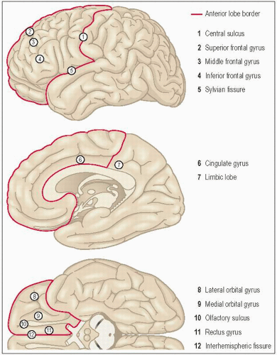
The anatomical boundaries of the frontal lobe (3.1) are demarcated posteriorly by the central sulcus of Rolando, mesially by the interhemispheric fissure, and inferiorly by the Sylvian fissure. The frontal lobe includes approximately one-third of the entire cortical surface of the brain. It is separated from the temporal lobe by the Sylvian fissure; medially it is separated from the limbic lobe by the cingulated gyrus; and separated from the parietal lobe by a line drawn from the marginal end of the central sulcus to the cingulate sulcus at the mesial border and by the central sulcus on the lateral border. The basal portion of the orbital surface is entirely in the frontal lobe. The lateral convexity contains the several gyri with the least variable gyral anatomy, including the precentral gyrus, superior, middle and inferior frontal gyrus. The medial aspect containing the cingulate gyrus and the basal surface is divided into gyrus rectus, as well as the medial and lateral orbital gyrus. The gyrus rectus is the medial most gyrus on the basal surface bordered by the olfactory sulcus on its lateral boundary.


Functional anatomy

The main functional regions of the cortex were defined by stimulation studies in the human and primate brain. They can be divided into primary motor areas, supplementary motor area (see Chapter 4 on Supplementary sensimotor
epilepsy), frontal eye field, frontal language areas (Broca’s area), negative motor areas, prefrontal cortex, and orbitofrontal and anterior medial areas (see Table 3.1).


3.1 The anatomical boundaries of the frontal lobe.

The primary motor areas include the posterior parts of the superior, middle and inferior frontal gyri and receive inputs from various sensorimotor regions. The main efferent output is the corticospinal tracts. This region is, as are many regions of the cortex, somatotopically organized. The frontal eye fields are located in the posterior part of the middle frontal gyrus in an area that immediately borders the primary motor cortex. Its main afferent input is from the occipital cortex and dorsal thalamus with its output projections to the preoccipital cortex and superior colliculus. The frontal language centre or Broca’s area is located in the region of the pars operculare and triangulare in the dominant frontal lobe. It is thought to have reciprocal connections to the receptive language centre located in the posterior part of the dominant temporal lobe (Wernecke’s area). Also, this region has connections to the primary tongue and laryngeal motor regions as well as the auditory cortex (Heschel’s gyrus) in the middle part of the dominant superior temporal gyrus as well as many other regions of the brain. The negative motor is found in the posterior part of the inferior frontal gyrus just in front of the primary motor cortex as well as in posterior aspect of the mesial frontal gyrus, which is almost continuous with the supplementary motor area in this region. The functional properties of the premotor cortex and orbitofrontal areas are less clearly defined. They include functions involved in
executive function, such as working memory and processing of sensory inputs of various modalities.


Table 3.1 Anatomy of the frontal lobe


Regions


Areas of cortex


Afferents


Efferents


Primary motor area


Posterior parts of the superior


Middle and inferior frontal gyri


Sensorimotor regions


Corticospinal tracts


Frontal language area (Broca’s area)


Pars operculare and triangulare


Posterior part of temporal lobe (Wernicke’s area)


Tongue and larynx motor, Wernicke’s area


Frontal eye field


Posterior part of the middle frontal gyrus bordering on primary motor cortex


Occipital cortex dorsal thalamus


Preoccipital cortex superior colliculus


Negative motor areas


Posterior part of inferior frontal gyrus and posterior mesial superior frontal gyrus


Prefrontal areas


No direct connections to spinal cord or primary motor areas


Seizure semiology

The cortical areas involved in the production of the seizure, the symptomatogenic zone, will determine the clinical manifestation of the seizure, i.e. seizure semiology. The knowledge of the initial symptomatogenic zone based on a patient’s clinical description of the seizure may often give some information on the localization of the region, where the epilepsy arises from or the epileptogenic zone. However, there are different regions of the cortex that can produce the same clinical manifestation of a seizure, as so many times the sensitivity of the seizure semiology to locate the epileptogenic zone is poor. Another issue to keep in mind is that the initial manifestation of the seizure might reflect propagation patterns of the seizure and not information on the epileptogenic zone, particularly as there are large regions of the cortex that are ‘silent’ in that when stimulated do not give rise to any clinical manifestations (Table 3.2).


Table 3.2 Seizure semiology of the frontal lobe


Symptogenic zones


Seizure type in the frontal lobe


Primary motor cortex


Focal clonic seizures (preserved consciousness)


Aura of ‘tightening of body parts’


Generalized tonic-clonic seizure


Frontal eye field


Head and or eye versive seizures


Anterior cingulate


Hypermotor seizures/complex motor seizures


Frontopolar


Hypermotor seizures/complex motor seizures


Frontal language area


Aphasic seizure


Negative motor area


Akinetic seizure


Seizures classified according to the semiological classification that can be seen in frontal lobe epilepsy are complex motor seizures. These seizures are defined as presenting with a variety of semipurposeful movements. At times they can involve more proximal musculature and are further classified as hypermotor seizures. The types of motor activity can include thrashing movements, and bicycling leg movements. The movements can be quite striking and often appear violent. These seizures tend to occur more often out of sleep. Other types of seizures include simple motor seizures originating from the primary motor cortex presenting as focal motor seizures affecting the contralateral limb of the facial muscles. These seizures can often occur in the presence of preserved awareness. Seizures arising from negative motor regions can present as a loss of tone or akinetic seizure. Seizures arising from frontal language areas can lead to a loss of speech with preserved awareness, which are called aphasic seizures. The frontal eye fields when involved in a seizure can lead to eye and or head versive movements (strong movements to the contralateral side). A variety of auras can also be seen in frontal lobe epilepsy; however, they are not of strong localization value. These auras include such descriptions as cephalic auras, abdominal auras and whole body sensations. Other descriptions such as ‘racing thoughts’ or a feeling of stiffening of a limb can also be seen in frontal lobe epilepsy.


Electroencephalography in frontal lobe epilepsy

Electroencephalography (EEG) findings during periods of time when no seizure is occurring in a patient, termed interictal EEG, can often be normal without any evidence of interictal epileptiform activity in up to 40% of patients. Epileptiform spike activity in frontal lobe epilepsy may not appear focal or restricted to the frontal lobe. Often spikes can appear lateralized to one hemisphere or even generalized. In some instances a leading spike can be seen preceding a more diffuse or generalized spike, which is termed secondary bilateral synchrony. When this finding is recognized it can indicate focal epilepsy as opposed to generalized epilepsy (see case 1).

When focal epileptiform spikes are seen, they are more likely to occur in the lateral frontal cortex and may be seen in about 50% of these cases. In the absence of a magnetic resonance imaging (MRI) lesion, focal epileptiform activity can often give some guidance for further testing or suggestions to specific modalities to further elucidate the epileptogenic zone.

EEG identified activity seen during a seizure, or ictal EEG, can vary in its utility in localization or even lateralizing the epilepsy (see case 2). At times, an electromyography artefact obscures the EEG so that no clear information is obtainable. This can be seen in 20% of the cases. At other times a more diffuse pattern can be seen but again not lending any further localization value. Localizable ictal EEG patterns are described in only 30-40% of cases with frontal lobe epilepsy. At times when the surface EEG shows a diffuse pattern an invasive EEG recording with subdural arrays can provide a more localized seizure onset. Focal scalp ictal onset patterns often consist of low amplitude fast activity, occasionally high amplitude spike or polyspike activity, or a leading focal spike/transient prior to more diffuse changes seen on EEG (see case 3).

Only gold members can continue reading. Log In or Register to continue

Stay updated, free articles. Join our Telegram channel

Aug 1, 2016 | Posted by in NEUROLOGY | Comments Off on Frontal Lobe Epilepsy

Full access? Get Clinical Tree

Get Clinical Tree app for offline access