General Principles of Electroencephalography
QUESTIONS
1. Which of the following electrodes is nonpolarizable?
A. Gold
B. Platinum
C. Silver
D. Silver-silver chloride
View Answer
1. (D): Electrodes become polarized as a result of accumulation of charges that may favor current flow in one direction over another. This is more likely with certain materials and with recording direct current (DC) potentials. Silver-silver chloride electrodes are nonpolarizable. (Fisch, pp. 23-24; Ebersole and Pedley 2003, pp. 50-54)
2. Using the international 10-20 electrode system, if the distance between the nasion and the inion is 38 cm, what would the distance from Fz to Pz be?
A. 15.2 cm
B. 19.0 cm
C. 27.8 cm
D. 30.2 cm
View Answer
2. (A): The international 10-20 system electrode system is based on the distance from the nasion to the inion. Fpz and Oz are measured at 10% of the distance respectively from the nasion and inion. The distance from Fz to Cz and from Cz to Pz is 20% of the total nasion-inion distance. So the distance from Fz to Pz will be 40% of 38 cm, that is, 15.2 cm. (Ebersole and Pedley 2003, pp. 72-75)
3. The following is true about the combinatorial nomenclature of the 10-10 system as compared to the international 10-20 system:
A. T7/T8 replace T3/T4
B. P7/P8 replace T5/T6
C. A and B
D. None of the above, there is no difference in electrode naming
View Answer
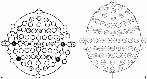
3. (C): The modified combinatorial nomenclature for the 10-10 electrode system uses the same landmarks as those of the 10-20 system for electrode placement. The 10-10 system expands the number of electrodes by adding new electrodes in between the 10-20 system electrodes. This is often used during long-term video-EEG monitoring where more accurate localization can be made. The number after the letter refers to the electrode position in relation to midline. In order to maintain this rule T3/T4 and T5/T6 electrodes had to be renamed. T7/T8 replace T3/T4 and P7/P8 replace T5/T6 (see Fig. 4.4). (Fisch, pp. 551-553; Abou-Khalil and Misulis 2006, pp. 12-15)
4. High-frequency filter of 30 Hz would most attenuate:
A. 15 Hz
B. 30 Hz
C. 55 Hz
D. 70 Hz
View Answer
4. (D): Using a 30-Hz high-frequency filter (low pass filter) will attenuate any frequency >30 Hz. Higher frequencies are more attenuated. In this case, the highest frequency is 70 Hz and will be the most affected by that filter setting. (Daube 2002, pp. 12-13)
5. Amplitude of scalp-recorded electroencephalogram (EEG) potentials depends on:
A. Intensity of the potential
B. Spatial orientation of the potential
C. Resistance of the structures
D. All of the above
View Answer
5. (D): The amplitude of a potential depends on its intensity, spatial orientation, and distance that separates it from the scalp. The resistance and capacitance of the structures between the generator of the potential and the scalp recording electrodes also affect inversely the amplitude. This is why scalp-recorded EEG is of lower amplitude than EEG activity recorded directly from the brain surface. (Fisch, pp. 14-16; Daube 2002, pp. 28-39)
6. Compared to near-field potentials, far-field potentials typically have:
A. Smaller scalp distribution
B. Wider scalp distribution
C. Higher voltage
D. Better localizing value
View Answer
6. (B): Scalp EEG signals record electrical activity originating from a generator through volume conduction. Near-field and far-field potentials respectively refer to potentials generated near and distant from the recording site. In the case of far-field potentials, the recording electrode captures a potential that has traveled through multiple heterogeneous media using volume conduction. As a result, the potential will have a wider scalp distribution and a lower voltage. (Wyllie, Gupta and Lachhwani 2005, pp. 142-143; Daube 2002, pp. 184-185)
7. Increasing the number of seconds displayed on the monitor:
A. Decreases spike amplitude
B. Decreases spike duration
C. Decreases spike frequency
D. Does not change spike amplitude, duration, or frequency
View Answer
7. (D): In analog or digital systems, increasing the paper speed or compressing the EEG recording will not have any impact on spike duration, amplitude, or frequency. Increasing the number of seconds displayed on the monitor may help better visualize focal slow activity, evolution of ictal discharges, and identification of long-interval periodic discharges. (Niedermeyer and Lopes Da Silva 1999, pp. 136-140)
8. Which of the following best describes the amplitude of the scalp potential of an EEG signal?
A. It is proportional to the solid angle of the cortical generator at the electrode site
B. It is inversely proportional to the solid angle of the cortical generator at the electrode site
C. It is proportional to the distance from the cortical generator to the recording electrode
D. None of the above
View Answer
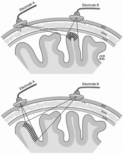
8. (A): The orientation of a cortical dipole in relation to the recording electrode is a major factor in determining the scalp electrical potential. This net potential is directly proportional to the cortical generator solid angle subtended by the recording electrode (see Fig. 4.5). (Wyllie, Gupta and Lachhwani 2005, pp. 144-145; Niedermeyer and Lopes Da Silva 1999, pp. 146-148)
9. The intracellular pathophysiologic mechanism of a focal epileptiform discharge is:
A. Action potential
B. Paroxysmal depolarizing shift
C. Presynaptic excitation
D. Hyperpolarization
View Answer
9. (B): The paroxysmal depolarizing shift (PDS) represents the intracellular electrophysiological correlate of focal epileptiform discharges such as sharp waves and spikes. It consists of abnormal synchronous activation of multiple neurons at the cellular level causing a wave of depolarization. It is primarily due to the activation of high-frequency fast sodium channel potentials. (Niedermeyer and Lopes Da Silva 1999, pp. 28-32; Daube 2002, pp. 57-59)
10. The ipsilateral earmontage shows prominent electrocardiogram (ECG) artifact greater on the left. Which of the following is the best strategy to minimize the artifact?
A. Switch to contralateral ear reference
B. Switch to left ear reference
C. Switch to right ear reference
D. Using a linked ear reference
View Answer
10. (D): ECG artifact represents a far-field potential recorded at the scalp. The orientation of the cardiac field produces opposite polarity potentials at each ear. When the ears are linked, the opposite polarities of the ECG artifact will cancel out (especially if the ECG potentials are equal in voltage) leading to a less contaminated reference. ECG artifact is more pronounced in obese patients, children, and patients with short necks. (Ebersole and Pedley 2003, pp. 275-276)
11. In a bipolar montage, all sharp waves will be displayed as a reversal of polarity.
A. True
B. False
View Answer
11. (B): Bipolar montages both longitudinal (commonly referred to as double banana) or transverse link sequential pairs of electrodes into channels. Recorded potentials affecting one electrode will produce reversal of polarity in channels having this electrode in common. If two adjacent electrodes inside the chain are equally and maximally affected, the channel containing the two electrodes will show cancellation, but reversal of polarity will be noted across that channel. Reversal of polarity is not noted if the recorded potential (in this case a sharp wave) maximally involves the electrode at the beginning or end of the chain (end of chain phenomenon). (Abou-Khalil and Misulis 2006, pp. 31-33)
12. Which thalamic nucleus does not project directly to the cortex?
A. Pulvinar
B. Reticular nucleus
C. Anterior nucleus
D. By definition, all thalamic nuclei project directly to the cortex
View Answer
12. (B): All of the thalamic nuclei project directly to the cortex except the reticular nucleus of the thalamus. This nucleus receives input from the cortex and sends its output to other thalamic nuclei. It plays a major role in thalamocortical connectivity and is thought to be primarily responsible for the electrical synchronization that occurs with seizures. (Fisch, pp. 10, 410-411)
13. Which of the following electrode pairs aremost sensitive respectively to vertical and horizontal eye movements?
A. T3/T4 and F7/F8
B. Fp1/Fp2 and F7/F8
C. F3/F4 and T3/T4
D. Fp1/Fp2 and T3/T4
View Answer
13. (B): Using the international 10-20 system, vertical eye movements are best seen in the frontopolar electrodes notably Fp1 and Fp2. Horizontal or lateral eye movements will be best recorded at F7 and F8 electrodes which are the nearest to the lateral side of the eyes. (Fisch, pp. 107-110)
14. During long-term video-EEG recording, which of the following material is preferably used to attach electrodes to the scalp?
A. Acetone
B. Collodion
C. Gorilla glue
D. Super glue
View Answer
14. (B): Several different techniques are used to attach electrodes to the scalp. In the case of long-term monitoring, the collodion technique offers a very stable recording with minimal artifact. In this technique, few drops of collodion are applied around the edge of the electrode. Stream of compressed air is used to dry the collodion. Adequate hyperventilation is needed for the safe use of collodion or acetone that removes collodion. (Fisch, pp. 20-21)
15. Which of the following dipoles contribute least to the scalp EEG?
A. Horizontal
B. Radial
C. Circular
D. Oblique
View Answer
15. (A): Every EEG potential has a dipole with negative and positive ends. The EEG is best able to record radial dipoles that are perpendicular to the crown of the gyri. These dipoles are also called vertical or perpendicular. For these dipoles, only one end of the dipole is recorded on the scalp electrodes while the other end is directed deep into the brain. (Fisch, pp. 87-90)
16. The maximal voltage of a sharply contoured activity at F3 is −70 μV and at Fz is −30 μV. What would the voltage be across the channel Fz-F3?
A. −40 μV
B. 40 μV
C. −100 μV
D. 100 μV
View Answer
16. (B): The voltage across the channel Fz-F3 is obtained by calculating the difference between the voltages at Fz and F3, i.e. (−30 μV) – (−70 μV) = 40 μV. (Abou-Khalil and Misulis 2006, pp. 30-38)
17. γ-Aminobutyric acid A (GABAA) receptors modulate:
A. Sodium channels
B. Potassium channels
C. Chloride channels
D. Calcium channels
View Answer
17. (C): Activation of GABAA receptors open chloride channels causing hyperpolarizing potentials. Therefore, GABAA receptors are responsible for fast synaptic inhibition. Evidence suggests that impaired GABAA receptor function is the pathological basis of some inherited and acquired epilepsies. (Wyllie, Gupta and Lachhwani 2005, p. 94)
18. What are the electrical charges of the cornea and tip of the tongue?
A. Cornea and tip of tongue are positively charged
B. Cornea and tip of tongue are negatively charged
C. Cornea is positively charged and tip of tongue is negatively charged
D. Cornea is negatively charged and tip of tongue is positively charged
View Answer
18. (C): Compared to the retina, the cornea is positively charged. This is illustrated when the EEG records eye closure. During eye closure, there is elevation of the eye (Bell’s phenomenon) causing a positive potential (or downward deflection) in the frontopolar electrodes. The tip of the tongue is negatively charged as compared to its base. This is illustrated when the EEG records chewing.With mastication, there is rhythmic elevation of the tip of the tongue causing upward sinusoidal waves mimicking frontal rhythmic activity. (Abou-Khalil and Misulis 2006, pp. 82-93)
19. The duration range of a sharp wave is:
A. 0-70 ms
B. 70-200 ms
C. 200-370 ms
D. It is not defined by duration
View Answer
19. (B): Spike and sharp waves are transient epileptiform discharges defined by their duration. The duration of spikes is <70 ms whereas that of a sharp wave ranges from 70 to 200 ms. (Abou-Khalil and Misulis 2006, pp. 48-49)
20. In the 10-20 international nomenclature, EEG activity from the F7 and F8 electrodes localizes to the:
A. Inferior frontal region
B. Anterior temporal region
C. Mesial frontal region
D. A or B
View Answer
20. (D): According to the international 10-20 system, the F7 and F8 electrodes lie over inferior frontal cortex, but most often record anterior temporal electrical activity. Depending on their field, they are referred to as inferior frontal or anterior temporal electrodes, and are sometimes referred to as the frontotemporal electrodes. One way to determine the source of their activity is to evaluate involvement of adjacent electrodes. For example, if T3 is also involved, F7 is likely recording anterior temporal activity, and if F3 is also involved, F7 is more likely to be recording inferior frontal activity. (Abou-Khalil and Misulis 2006, pp. 10-12)
21. In the intensive care unit (ICU), a ground loop can be avoided by:
A. Using multiple patient grounds
B. Using a long extension cord
C. Connecting each piece of equipment to a separate outlet ground
D. Using one common ground
View Answer
21. (D): A ground loop occurs when two or more instruments are attached to the patient and their ground levels are unequal. In that case, any leakage current from one instrument with higher voltage ground level may travel through the patient and then flow to the ground electrode of another instrument with a lower voltage creating ground a loop circle. This may produce a circuit of current flow referred to a ground loop, which increases the risk of patient electrocution. To avoid a ground loop circuit, one common ground is advisable especially in environments like the ICU, where multiple electrical equipments are attached to the patient. (Fisch, p. 71)
22. Tetrodotoxin is a:
A. Sodium channel blocker
B. Calcium channel blocker
C. Potassium channel blocker
D. GABA receptor blocker
View Answer
22. (A): Tetrodotoxin (TTX) is a nonprotein organic compound and one of the strongest paralytic toxins. It is a specific irreversible blocker of voltage-gated sodium (Na+) channels. It has been extremely valuable in Na+ channel identification and characterization in animal epilepsy models as well as in antiepileptic drug mechanisms. Similarly valuable was tetraethyl ammonia (TEA), a selective potassium channel blocker. (Wyllie, Gupta and Lachhwani 2005, pp. 75, 78-79)
23. What is the mechanism of action of baclofen, a drug than lowers seizure threshold?
A. GABAA agonist
B. GABAA antagonist
C. GABAB agonist
D. GABAB antagonist
View Answer
23. (C): Baclofen is a GABAB agonist medication commonly used for spasticity. GABAB is a G-protein-coupled receptor that can open potassium or close calcium channels. Its receptors are presynaptic and postsynaptic. When activated, it modulates T-calcium channels and intrinsic bursting activities of the relay cell causing seizures. Benzodiazepines are GABAA agonists. (Wyllie, Gupta and Lachhwani 2005, pp. 94-95)
24. Scalp-recorded electrical activity arises from:
A. Action potentials generated in the deep layers of the cortex
B. Action potentials generated in the superficial layers of the cortex
C. Postsynaptic potentials generated in the superficial layers of the cortex
D. Postsynaptic potentials generated in the deep layers of the cortex
View Answer
24. (D): Postsynaptic potentials and not action potentials contribute to the scalp-recorded electrical activity. This is because postsynaptic potentials are of long duration ranging from 10s to 100s of milliseconds. They involve a large area of the membrane surface and occur simultaneously in thousands of cortical cells arranged perpendicularly to the cortical surface. (Daube 2002, p. 61)
25. In order to record a spike on scalp EEG, the minimal surface area of cortex involved is:
A. 0.6 mm2
B. 6 mm2
C. 0.6 cm2
D. 6 cm2
View Answer
25. (D): The cortical surface involved is at least 6 cm2 for a potential to be detected on scalp EEG. This is one reason why many simple partial seizures involving a small cortical area are often missed on scalp EEG. (Fisch, pp. 13-16)
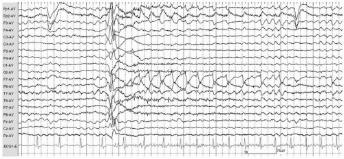
26. The EEG recording shown in Figure 4.1 ismost consistent with:
A. Roving eye movements
B. Ictal nystagmus
C. Eye flutter
D. Breach rhythm
View Answer
26. (B): This EEG tracing is displayed in a referential average montage. After ictal onset (with the high voltage sharply contoured wave) there is rhythmic activity of opposite polarity at F7 and F8. Each positive deflection at F7 is preceded by a small spike representing lateral rectus muscle activity. The pattern is likely due to horizontal left-beating nystagmus. Lateral eye movements are best recorded at the electrodes closest to the eyes. The reversal of polarity is due to the movement of the positively charged cornea toward one electrode and away from the other electrode. Roving eye movements would be of much slower frequency. Eye flutter produces vertical eye movements best seen in the frontopolar electrodes. (Ebersole and Pedley 2003, pp. 273-275)
27. Bancaud phenomenon is a normal EEG variant.
A. True
B. False
 Figure 4.1.
Stay updated, free articles. Join our Telegram channel
Full access? Get Clinical Tree
 Get Clinical Tree app for offline access

|


