Parameter
Range
CSF glucose
2.7–4.2 (mmol/L)
Blood glucose
3.3–5.5 (mmol/L)
Glucose CSF/blood ratio
0.61–0.89
(Cave: CSF/serum ratio may be lower than 0.5 if serum glucose concentration is >100 mg/dL) (see Fig. 7.1)
7.1.4 Interpretation
CSF/blood ratios below 0.5 are indicative of bacterial CNS infections caused by organisms such as streptococcus, meningococcus, mycobacteria, and funguses. During meningeal blastomatosis, CSF levels of glucose may also drop below this ratio of 0.5. However, in cases with increased serum glucose concentrations, the normal CSF/blood glucose ratio may drop up to below 0.4 (see Fig. 7.1a, b), and therefore an apparently decreased CSF/serum ratio may be due to a diabetic dysmetabolism which requires adjustment to avoid a misinterpretation (Hegen et al. 2014; Leen et al. 2012). According to a recent study, cutoff values for normal CSF/SGlu ratio defined as the 5th percentile were 0.5 for patients with serum glucose concentrations <100 mg/dL, 0.4 for those with a glucose level of 100–149 mg/dL, and 0.3 for serum glucose concentrations ≥150 mg/dL (Hegen et al. 2014).


Fig. 7.1
(a) Relationship between blood glucose (mmol/L) and CSF/blood glucose ratio (Leen et al. 2012) (b) and relationship between serum glucose (mg/dL) and CSF/serum glucose ratio (Hegen et al. 2014). Due to the inverse correlation, cutoff levels for normal CSF/serum glucose ratio require adjustment to the glucose concentration in blood or serum, respectively
7.2 Lactate
Lactate in human brain is produced by glial cells, mainly astrocytes, and it is aerobically metabolized by neurons (Brooks 2002). Lactate in CSF is derived from the CNS and from blood. However, changes in blood concentration do not affect the CSF concentration, since the lactate transporter at the blood-brain barrier is saturated (Barros and Deitmer 2010). Therefore, when evaluating CSF concentration of lactate, knowledge of the blood level is not required.
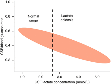
Lactate occurs in two optical isomers, l-lactate and d-lactate. l-Lactate concentration predominates with up to 99 %; thus it is of major importance in routine CSF analysis and d-lactate has no established role in routine CSF diagnostics. l-Lactate represents the end product of the anaerobic glucose metabolism, so it can replace glucose analysis (Fig. 7.2). Elevation of lactate indicates hyperlactatemia or lactic acidosis which is an important diagnostic aspect in intensive care units.


Fig. 7.2
The inverse relationship between CSF/blood glucose ratio and CSF lactate concentration explaining why CSF lactate as a single parameter is equally informative as CSF/blood ratio of glucose
7.2.1 Preanalytics
Stability of lactate in CSF strongly depends on the number of leukocytes, red blood cells and pathogens. Red cells in CSF cause significant increases in lactate concentrations, more so when exposed to air. This has to be considered when interpreting lactate in CSF contaminated by blood (Venkatesh et al. 2003). Lactate is stable at room temperature for up to 24 h independent of CSF leukocyte count without addition of sodium fluoride (Dujmovic and Deisenhammer 2010; Thomas 2005).
Stay updated, free articles. Join our Telegram channel

Full access? Get Clinical Tree


