Associations/Risk Factors
Apart from blunt trauma and age >60 years, risk factors for CSDH include coagulopathies, for example related to liver disease, iatrogenic therapeutic anticoagulation or alcohol abuse; recurrent falls, for example in patients with dementia syndromes or with hemiplegia as a result of previous stroke; any condition causing significant cerebral atrophy (alcoholism, dementia etc.) and patients with shunts, for example a ventriculoperitoneal shunt for normal pressure hydrocephalus (see Chapter 37). Anticoagulation therapy also increases the risk for an ASDH.
Pathology/Pathogenesis
ASDH are usually associated with severe impact damage and primary injury to the brain. It can arise either from oozing/accumulation around an area of laceration to the brain parenchyma, usually in the frontal or temporal region or due to rupture of a bridging vein from acceleration–deceleration during rapid movement of the head. In the latter case, particularly in cases where the bridging veins are stretched due to cerebral atrophy and therefore more susceptible to injury with lesser degrees of force, the underlying brain injury may be mild and patients’ presentation may mimic that of an acute extradural haematoma with a lucid interval followed by a rapid deterioration. Rupture of a cortical artery may also result in ASDH.
History
ASDH, usually subsequent to trauma, for example falls or road traffic accidents, can mimic extradural haematoma in its presentation with a lucid interval prior to deterioration. In younger patients, ASDH is typically associated with high-velocity blunt head trauma and patients are often comatose from the outset. It can also present with a more gradual deterioration in GCS with haematoma expansion, headache, nausea and vomiting. Seizures may occur in the posttraumatic period.
Diagnosis of CSDH on average occurs 4–5 weeks following trauma. CSDH patients can present insidiously with symptoms mimicking TIA, headaches, confusion and cognitive decline, nausea and vomiting, change in personality and episodes of speech arrest or dysfunction (with dominant hemisphere lesions). Fluctuation in symptoms may be marked. Patients may complain of weakness, usually contralateral to the side of the haematoma.
Examination
Findings associated with ASDH may include the following (also see Chapter 30):
- Deterioration in conscious level (GCS) as above.
- Dilated, unreactive pupil ipsilateral to the haematoma.
- Focal neurological deficits, for example dysphasia and sensory disturbance, and long tract signs, for example hemiparesis usually contralateral to the side of haematoma.
Findings associated with CSDH may include the following:
- Reduced level of consciousness, focal neurological deficits as above, reflex asymmetry and gait disturbance.
- Evidence of dysphasia (may be fluctuating) with dominant hemisphere collection.
- Reduced scores on mini-mental state examination in patients with CSDH and associated cognitive decline (a treatable cause of dementia).
Investigations
- Blood tests: FBC, U & E (particularly sodium level check) and clotting parameters (correct any coagulopathy).
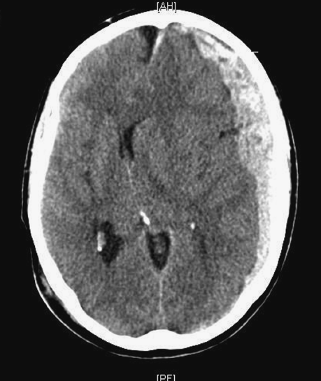
- CT: Crescentic blood collection (concave towards brain surface) with varying attenuation depending on the clot age. An acute collection (Figure 33.1) will be hyperdense, subacute isodense (Figure 33.2) and chronic hypodense (similar to the density of CSF) (Figure 33.3) in comparison to the density of brain parenchyma. Some collections may have mixed density, for example due to a new haemorrhage into an old collection, the so-called ‘acute on chronic SDH’.
Figure 33.1 CT demonstrates a large hyperdense ASDH over frontal, temporal and parietal convexity with significant mass effect, i.e. midline shift and effacement of ventricles. Note the characteristic crescentic shape of the clot.