Localization
Percentage (%)
Liver
68
Lung
23
Kidney
2
Brain
2
Bone
2
Striated muscle
1
Spleen
1
Multiple sites
1
E. granulosus infection of the CNS has been estimated at below 2 % (Schantz 1972) and may be primary, often presenting as solitary parenchymal cysts, while intraventricular and meningeal locations are most exceptional (Arana Iñiguez et al. 1951). Secondary involvement is due to cyst rupture or hydatid fluid dissemination from a distant organ. CNS echinococcosis is more frequent in children up to 15 years of age with an estimated prevalence rate of 50–75 % (Dew 1955; Slim et al. 1971). A series of cerebral hydatid cysts in children describing diagnostic difficulties, surgical treatment, and possible complications have been reported (Krajewski and Stelmasiak 1991; Ersahin et al. 1993; Ozek 1994; Peter et al. 1994; Turgut 2002). Signs and symptoms include increased intracranial pressure, headache, vomiting, papilledema, and hemiparesis as well as occasional seizures, while meningeal or cranial nerve involvement is most unusual (Arana Iñiguez 1978; Kammerer 1993).
Epidemiology
Hydatidosis in South America
Hydatid disease is endemic in cattle- and sheep-raising regions of the world such as Central Europe, the Mediterranean countries (Tunisia, Spain), the Middle East (Turkey, Lebanon, Emirates), South America (Argentina, Peru, Uruguay, Brazil, Chile, and Bolivia), Australia, New Zealand, India, and South Africa (Carrea et al. 1975). South America is one of the regions of the world most affected by cystic echinococcosis.
Three of the four species of Echinococcus, E. granulosus, E. vogeli, and E. oligarthrus, have been described in South America (Fig. 3.1). However, E. granulosus presents transmission cycles in domestic animals, being sheep the principal intermediate hosts in this region. The reason for this fact is the great concentration of sheep in this part of the continent and the close relationship between this intermediate host and the definitive host, the dog. There are now approximately 65 million sheep in South America, out of which 63 million (97 %) are found in Argentina, Brazil, Chile, Peru, and Uruguay. These data turn nearly 9 % of the South American territory into a sheep-raising area (Organización Panamericana de la Salud [OPS] 2004). However, in certain regions, various animal species such as pigs, goats, and camelids have gained importance as intermediate hosts. In South America, a few described human cases of alveolar echinococcosis were due to E. patagonicus and E. oligarthrus (Taratuto and Venturiello 1997). The prevalence rate of infection in the asymptomatic human population obtained from ultrasonographic (US) screenings was 1.6 % in Tacuarembó, Uruguay; 1.6 % in Florida, Uruguay; 3.6 % in Durazno, Uruguay; and 5.1 % in Vichaycocha, Peru. In Argentina, the prevalence rate was 5.5 % in Rio Negro province and 14.2 % in Loncopue, Neuquen province (OPS 2004). Successful control programs based on systematic dog deparasitation with praziquantel have been developed in Uruguay, Chile, and Argentina.
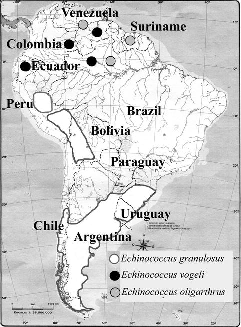
Fig. 3.1
Distribution of Echinococcus granulosus, E. vogeli, and E. oligarthrus in South America (Modified from OPS (Organización Panamericana de la Salud) 2004; with permission)
In general, hydatid disease of the CNS is rare, with a reported incidence of 0.9–2.1 % of all cases of hydatidosis (Tamer et al. 2004). A differential diagnosis with other diseases such as abscesses, neoplasias, and other parasitoses such as coenurosis and congenital cysts should be determined (Mahadevan et al. 2011).
Hydatidosis in Argentina
In Argentina, E. granulosus is the most prevalent zoonotic helminth. It has been estimated that approximately 30 % of the Argentine territory harbors the domestic life cycle of E. granulosus. The provinces where sheep, goat, and cattle rearing is quite common are the ones with the highest prevalence rates of infection. In particular, Neuquén is one of the provinces with the highest incidence (OPS 2004).
Although E. granulosus is widespread all over Argentina and hydatid disease is considered endemic in this country, the infection has a nonhomogeneous geographic distribution (Fig. 3.2). The contribution of each kind of livestock (cattle, pig, sheep, etc.) to disease transmission depends on the region: for example, while sheep are the principal intermediate hosts in the Patagonian and Mesopotamic region, cattle are the prevalent hosts in the Pampas and northern regions of the country (Denegri et al. 2002). Particularly in the southeastern region of Buenos Aires province, echinococcosis is considered an important human and veterinary health problem, cattle being the main livestock involved in the transmission of this disease (Andresiuk et al. 2009). In a case-control study in Argentina, one of the risk factors for cystic echinococcosis was human exposure to a great number of dogs during the first years of life (Larrieu et al. 2002). In a survey of asymptomatic patients, Coltorti et al. (1988) found that 47 of 5,839 patients (80 %) were seropositive for E. granulosus antigens. These authors used antibodies against E. granulosus arc 5 antigens by the double diffusion arc 5 test (Coltorti et al. 1988).
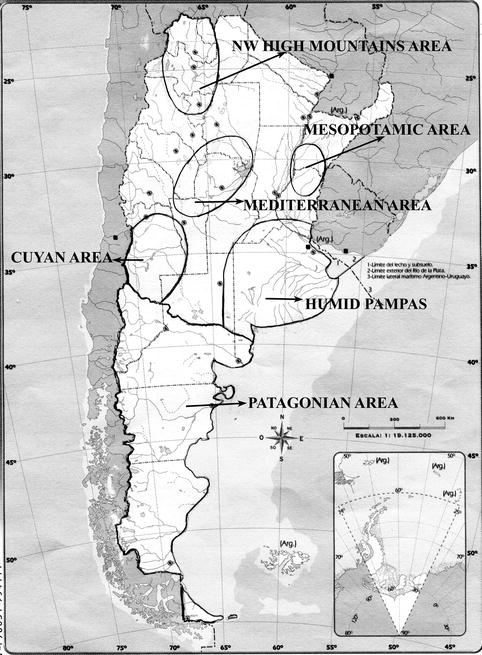
Fig. 3.2
Major endemic foci of echinococcosis in Argentina (Modified from OPS (Organización Panamericana de la Salud) 2004; with permission)
Argentine medicine has greatly contributed to the comprehension of hydatid cysts of the CNS. In Buenos Aires in 1889, Dr. Alejandro Castro performed a surgical operation for a cerebral hydatid cyst and introduced the new principles of surgical asepsis that had been previously developed in Europe. In 1895, Dr. Alejandro Posadas was the first surgeon to successfully treat hydatid cysts of the CNS in Argentina. In 1929, Dr. Ernesto Dowling performed his so-called “cyst delivery” method. The hydatid cyst is surgically removed intact. This technique (Dowling-Orlando technique) is mandatory in order to avoid further dissemination of the scolices (Obrador Alcalde 1964). In Buenos Aires, during the 1933–1974 period covering almost 42 years, 29 brain hydatid cysts and one of the spinal cord out of 35 pediatric cases of hydatidosis of the CNS were operated. No surgical mortality was reported in these cases (Carrea et al. 1975). The 20 patients who had their cysts removed unruptured with the Dowling technique (Dowling and Orlando 1939) remained alive, and only two patients suffered blindness as a sequela (Carrea et al. 1975). In Buenos Aires, in 1980, three patients (two adults and one child) were admitted to hospital with various neurological signs and symptoms. Although there was no evidence of cysts in the liver or lungs, hydatid disease was only suspected in the child, since he came from an endemic rural area in Buenos Aires province. A high degree of accuracy in identifying multiple supratentorial cysts by computed tomography (CT) scanning was obtained in all the cases, and the patients were followed by CT scans after surgery (Insausti et al. 1980). In Buenos Aires in 1998, Ure and Maegli reported a case of an adult patient born in Santiago del Estero province, who was admitted to a psychiatric hospital after surgery performed in Buenos Aires 1 year before (Ure and Maegli 1998). He suffered from alcoholism, diabetes mellitus, and Chagas trypanosomiasis. He had lived on a farm for several years and then moved to Buenos Aires. He presented with excruciating headache, vomiting, papilledema, left hemiparesis, somatic sensitivity in the left leg, slow mental activity, and urinary incontinence. A CT scan revealed a large right frontal cystic mass surrounded by a circular calcification. The mass turned out to be a hydatid cyst, which was opened and treated with formaldehyde. No extracerebral localizations were observed. Five years later, new CT scans revealed the bilateral and diffuse extension of the hydatid process with several small new cystic lesions unable to be treated by surgical reoperation. The patient died 2 months later (Ure and Maegli 1998).
In 2004, a case of a temporal hydatid cyst in a child was reported. The diagnosis was considered preoperatively on the basis of magnetic resonance imaging and magnetic resonance spectroscopy (MRS) findings. The MRS pattern appeared to be different from that seen in other cystic lesions of the CNS. Lactate, alanine, acetate, and a large pyruvate peak were observed. The cyst was surgically removed (Tamer et al. 2004).
Hydatidosis in Brazil
In Brazil, Echinococcus is widespread in an area of approximately 100,000 km2, which is close to the Uruguayan and Argentinean borders. Several studies have shown that 14–50 % of farm dogs were infected with E. granulosus (de la Rue 1997). Particularly in the state of Rio Grande do Sul in southern Brazil as well as in other countries in South America, the habit of feeding sheep raw offal to dogs perpetuates the parasite’s life cycle. The etiological agent found in dogs and other wild animals is predominantly E. granulosus.
In 1983, the first serological survey for echinococcosis was performed in seven municipal states near the border with Uruguay. A prevalence of 8.06/1,000 population was observed (OPS 2004). In 1999, another serological survey was conducted in 5 % of the total human population in rural areas of 18 municipal states of Brazil. The highest prevalence was observed in Municipio de Barra do Quaraí (89.44 %) and the lowest in Piratini (8.82 %) (Souza et al. 1999). To our knowledge, there have been no reported cases of hydatidosis of the CNS in the Brazilian literature.
Hydatidosis in Chile
In Chile, several serological surveys have been conducted throughout the country to document the prevalence of human echinococcosis from 1988 to 1997 (Nourbakhsh et al. 2010). The prevalence of human cases in apparently healthy people may reach up to 754.6/100,000 population in some regions of the country (Apt et al. 2000). Another serological survey reviewed 60,790 individuals and found that 82 had positive serological findings, resulting in a total incidence of 136/100,000 population. The calculated incidence rates for urban and rural regions were 87/100,000 and 241/100,000, respectively (Schenone et al. 1999). Pursuant to National Resolution N° 712/2000, echinococcosis is a disease of mandatory notification (OPS 2004) in Chile. Human echinococcosis is a prevalent disease in Chile with an incidence rate of 2–2.5/100,000 population. Considering the existing subdiagnoses, the incidence of this disease has been reported to be 10/100,000 (OPS 2004). In Chile, dogs are the main definitive hosts for E. granulosus. Between 1977 and 1979, 71 % of dogs from the XII region were found to be infected with Echinococcus In 1984, after the control program, this prevalence decreased to 1.5 % (OPS 2004).
Between 1997 and 2000, of 15 patients studied at Dr. L. Calvo Mackenna Hospital in Santiago de Chile, only 2 had cerebral hydatid cysts, whereas the rest had other cyst localizations (Hauck et al. 2003). More recently, in March 2012, four cases of cysts located in the brain were reported. Three of the patients were admitted with intracranial hypertension, whereas the fourth one presented with left hemiparesis. Imaging studies of all intracerebral cysts showed features compatible with hydatidosis in all cases. All lesions were completely removed surgically, and the pathological study of the excised piece confirmed the diagnosis of hydatidosis (Tapia et al. 2012).
Related posts:
 Hydatidosis of the Central Nervous System in Mediterranean Countries and the Middle East
 Historical Aspects of Hydatidosis of the Central Nervous System
 Surgical Treatment of Cerebral Hydatidosis
 Alveolar Hydatidosis of the Central Nervous System
 Complications of Spinal Hydatidosis
 Surgical Treatment of Spinal Hydatidosis
Stay updated, free articles. Join our Telegram channel

Full access? Get Clinical Tree


