Infants:
- Abnormal head growth with ‘crossing centiles’ on growth chart.
- Irritability, vomiting, apnoeic episodes and developmental delay.
Children and younger adults:
- Symptoms of raised intracranial pressure—headaches (typically waking from sleep in early morning, relieved on vomiting and worse on coughing/straining and lying down), nausea and vomiting and visual deterioration.
- Seizures are a relatively uncommon presenting feature but can be confused with ‘hydrocephalic attacks’—abrupt collapse with loss of consciousness due to abrupt rise in ICP. Recovery of consciousness can occur, for example due to transient relief of obstruction.
- More slowly progressive cases may not have headache, only visual failure due to occult optic atrophy and/or disturbance of gait (ataxia) and cognition (e.g. decline in school performance) and urinary incontinence.
Elderly:
- Triad of symptoms of NPH: Gait disturbance, dementia and urinary incontinence.
Shunt infection:
- If a shunt has been inserted within the last year there is a risk of it being infected.
- Ask about history of fever, abdominal pain and redness (erythema) along the course of shunt as it passes subcutaneously.
Examination
Infants:
- Head circumference over 98th centile; tense or full fontanelle; dilated scalp veins.
- Loss of upgaze ‘sunsetting’ (usually in obstructive hydrocephalus) due to downward pressure on tectum.
Children and younger adults:
- Signs of raised intracranial pressure: Decreased level of consciousness/coma, papilloedema and may have loss of upgaze; bilateral VI nerve palsies (false localising sign).
- Gait ataxia.
- Visual acuity, visual fields and fundoscopic findings should be documented in any patient with hydrocephalus or suspected shunt failure.
- Examine for evidence of an existing shunt system, the valve may be felt as a palpable lump under the scalp either on the top of the head behind hairline or a few centimetres above and behind the ear.
- Most shunt valves have a ‘reservoir’ that can be depressed and felt to refill if shunt is working. The shunt catheter may be palpated as it passes subcutaneously from scalp incision to abdominal incision. Look for any signs of infection of these incisions.
Elderly:
- Usually do not have signs of raised ICP. Typically gait ataxia and cognitive impairment only.
Investigations
CT head scan:
- Look for enlargement of ventricles (Evan’s ratio (frontal horn width/maximum biparietal diameter) >30%). Pattern of ventricular enlargement helps identify level of obstruction (e.g. in aqueduct stenosis, the IV ventricle is small and the lateral and III ventricles enlarged).
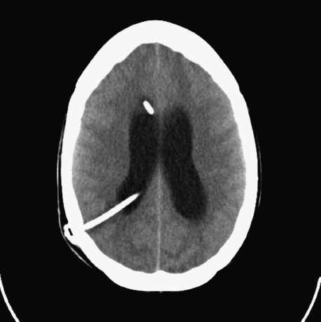
- The VP shunt catheter(s) are visible as a white (radio-opaque) linear structure (Figure 37.1).
Figure 37.1 CT head scan showing typical enlargement of ventricles. Note the ventricular shunt catheter visible as a radioopaque structure passing into the occipital horn of the right lateral ventricle and part of the proximal catheter of a separate ventricular access device (also known as an Ommaya reservoir) passing into the right frontal horn.
MRI scan:
- Can identify structural abnormalities causing obstructive hydrocephalus (e.g. tumours).
Shunt series:
- Skull AP and lateral, chest and abdominal X-rays.
- To assess continuity of VP shunt system: look for breaks in tubing and/or migration of catheters.
Ultrasound:
- Frequently used in infants with open fontanelle. Useful to monitor ventricular size.
ICP monitoring:
- Used when imaging is equivocal to determine whether ICP is raised.
Lumbar infusion study:
- Lumbar puncture is contraindicated in obstructive hydrocephalus.
- In communicating hydrocephalus, a one off ICP reading with a manometer can assist diagnosis, with drainage of CSF providing temporary symptomatic relief.
- An infusion study is a more sophisticated test measuring dynamic changes in ICP in response to infusion of artificial CSF into the lumbar subarachnoid space; it can be of use in the diagnosis of NPH or can help diagnose shunt blockage.
CSF drainage tests:
- Used in the diagnosis of NPH. Objective testing of gait and cognition before and after large-volume CSF drainage via a lumbar drain over a few days. Improvement in either gait or cognition correlates strongly with shunt responsiveness.
Neuropsychological testing:
- Can be used to monitor children or adults with hydrocephalus. A decline in cognitive performance may indicate shunt malfunction.
Management
Medical Treatment
- Hydrocephalus remains a surgically treated condition.
- Acetazolamide reduces CSF production and can be given as a temporising measure.
- Serial lumbar punctures may temporise hydrocephalus in cases of communicating hydrocephalus.
- If a deteriorating patient with known hydrocephalus has a ventricular access device (a ventricular catheter connected to a blind ending subcutaneous reservoir, usually located in the frontal region and separate from the shunt system) (Figure 37.1), this can be accessed using strict asepsis with a 25G needle to remove CSF in order to temporarily reduce ICP.
Surgical Treatment
Obstructive Hydrocephalus
Removal of cause:
- In some cases, such as tumours or colloid cysts, removal of the obstructing lesion can re-establish CSF flow.
Endoscopic third ventriculostomy (ETV):
- An operation in which a hole is made in the floor of the third ventricle into the subarachnoid space below it, allowing the obstruction (in the cerebral aqueduct or posterior fossa) to be ‘bypassed’. Successful in around 60% of cases.
Ventriculo-peritoneal (VP) shunt (Figure 37.1):
- A system for draining CSF from the ventricle to the peritoneal cavity, where it is absorbed.
- Three components: a ventricular catheter, which usually lies in the frontal or occipital horn of the lateral ventricle, connected to a valve (usually with an integral reservoir) and a distal catheter, which passes subcutaneously, draining the CSF (when the valve opens) to the peritoneal cavity.
- Other sites used occasionally for distal catheter drainage include the pleural cavity (ventriculo-pleural shunt) and the right atrium, via the internal jugular vein (a ventriculo-atrial (VA) shunt).
- Many different shunt valves are available. Most are differential pressure valves, opening at a fixed pressure. Programmable valves allow adjustment of the valve opening pressure.
Communicating Hydrocephalus and NPH
- These types of hydrocephalus are usually treated with a VP shunt as described above.
- The lumbar subarachnoid space is sometimes used as an alternative to ventricular catheter placement in communicating hydrocephalus, and is known as a lumbo-peritoneal (LP) shunt.
Complications
- Neither VP shunts nor ETV offer permanent cure of the condition; both may block leading to recurrent symptoms of hydrocephalus requiring revision surgery. Untreated hydrocephalus is often fatal.
- Delayed treatment can lead to permanent visual loss or significant cognitive impairment.
- Epilepsy can arise either due to underlying conditions or surgical intervention to treat hydrocephalus.
Shunts
Overdrainage:
- Shunts may drain too much of CSF causing low pressure symptoms such as headaches (typically relieved by lying down) and vomiting; in more severe cases, they can lead to collapse of ventricle that may cause tearing of bridging veins on the brain surface leading to subdural haematoma formation.
Infection:
- Rarely occurs more than 1 year after the shunt insertion (exception: VA shunts, which can become infected with any bacteraemia).
- Clinical signs include meningism and erythema along the line of distal catheter; confirm diagnosis with CSF culture from shunt reservoir.
- Requires shunt removal in most cases.
- Infected VA shunts can lead to renal failure due to shunt nephritis and pulmonary hypertension due to microemboli.
Blockage:
- Can occur at any time after shunt insertion, sometimes many decades later.
- Patients present with recurrent symptoms and signs of hydrocephalus.
Migration, disconnection, breakage of shunt catheters:
- Can be a cause of shunt failure. Look for evidence of loss of continuity of tubing on shunt series.
ETV
Blockage:
- Can still occur following ETV, often many years later and can be associated with fatality if not recognised. Clinical features of recurrent hydrocephalus.
Prognosis
Untreated:
- Fatal in approximately 80% of cases. Sixty per cent of survivors left with significant learning disability.
- Twenty per cent of survivors are blind.
Treated:
- Risk of mortality from shunt-related problems is 1–3% per year.
- With treatment, patients can expect to undergo regular shunt revisions.
- Infants have a 50% shunt failure rate within 1 year. Older children and adults on average undergo two shunt revisions every 10 years.
- Cognitive outcome is dictated by the presence of associated underlying conditions, although repeated episodes of raised ICP due to shunt complications or the development of an associated seizure disorder can have an adverse effect on cognition.
- Raised ICP due to hydrocephalus can lead to progressive optic atrophy with visual failure.
Differential Diagnosis (Table 37.1)
Table 37.1 Differential diagnoses of ventricular enlargement and of VP shunt failure.
| Ventricular enlargement | |
| External hydrocephalus |
|
| Ex vacuo ventricular dilatation |
|
| Arrested/compensated hydrocephalus |
|
| Hydranencephaly |
|
| VP shunt failure | |
| |
| Remember: The consequences of missing shunt failure are severe, so this diagnosis should be actively excluded. | |
< div class='tao-gold-member'>


