Hypothalamus, limbic system and olfactory system
In order to survive, there must be continual biochemical and physiological adaptations to preserve the internal environment of the body (homeostasis). Interoceptor signals from the internal organs and body fluids initiate homeostatic responses; consequently, the internal physical and chemical environment remains balanced and stable. The hypothalamus is the structure responsible for this task.
Exteroceptive information from the outside world dictates behavioural responses, in order to achieve individual ‘homeostasis’ within the physical and social environment. Behaviour is relatively simple and stereotyped in lower animals and directed to satisfying the drives of thirst, hunger, sex and defence, in instinctive repertoires. The limbic system, which is strongly connected to the hypothalamus, is essential for this adaptive behaviour, which includes the ability to learn new responses based on previous experience (memory). The complex and non-stereotyped behaviour of humans is an attempt to preserve the individual within the physical landscape but also within a changing social environment (individual homeostasis). The association areas of the neocortex are capable of analysing exteroceptive information from the environment and other individuals, enabling adaptive personal and social responses. These phylogenetically more recent structures are partly connected to the limbic system.
As a result, the hypothalamus, limbic system and association neocortices act as interfaces in a hierarchical fashion between the internal structure of the individual and the environment. A reminder of this evolutionary ascent in humans is the olfactory system: vital for sensing the environment in lower animals, overwhelmed by visuospatial dominance in humans and intimately related to the limbic system.
Hypothalamus
Topographical anatomy of the hypothalamus
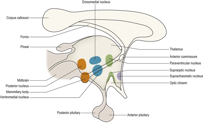
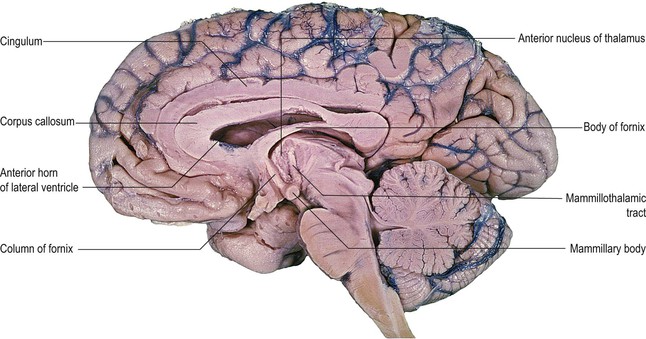
The hypothalamus is the most ventral part of the diencephalon, lying beneath the thalamus and ventromedial to the subthalamus (Fig. 16.1, see also Figs 12.1–12.3). It forms the floor and the lower part of the lateral wall of the third ventricle, below the hypothalamic sulcus (Fig. 12.2). On the base of the brain, parts of the hypothalamus can be seen occupying the small area circumscribed by the crura cerebri, optic chiasm and optic tracts (Fig. 12.1). Between the rostral limits of the two crura cerebri, on either side of the midline, lie two distinct, rounded eminences, the mammillary bodies, which contain the hypothalamic mammillary nuclei. In the midline, immediately caudal to the optic chiasm, lies a small elevated area known as the tuber cinereum, from the apex of which extends the thin infundibulum (infundibular process), or pituitary stalk. This is attached to the pituitary gland (hypophysis), a pea-sized structure which lies within the sella turcica of the sphenoid bone. The pituitary gland consists of two major, cytologically distinct, parts: the posterior pituitary or neurohypophysis and the anterior pituitary or adenohypophysis (Figs 16.2, 16.3). The posterior pituitary is a neuronal structure, being an expansion of the distal part of the infundibulum. The anterior pituitary is not neural in origin. The two parts are, however, closely linked by the pituitary (hypophyseal) portal system of vessels (Fig. 16.3), which are derived from the superior hypophyseal artery. Releasing factors, which are synthesised in the hypothalamus, pass to the adenohypophysis through these vessels to control the release of anterior pituitary hormones.
The hypothalamus is able to integrate interoceptive signals from internal organs and fluid-filled cavities and make appropriate adjustments to the internal environment by virtue of its input and output systems.
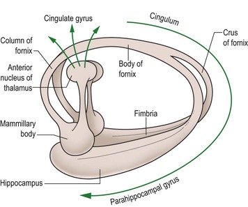
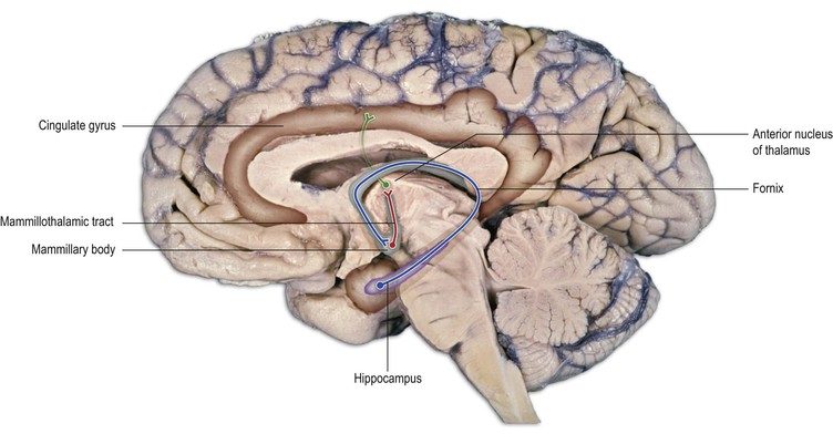
Input to the hypothalamus is both circulatory and neural in origin (Fig. 16.4). The circulating blood provides physical (temperature, osmolality), chemical (blood glucose, acid–base state) and hormonal signals of the state of the body, its growth and development and its readiness for action (e.g. sex, suckling, defence, escape, etc.). Neural signals come from a number of sources. The largest input originates from limbic structures, the hippocampus and the amygdala. Fibres of hippocampal origin constitute the fornix, a large component of which terminates in the medial mammillary nucleus within the mammillary body (Figs 16.5–16.7). Fibres from the amygdala to the hypothalamus run in the stria terminalis. The nucleus solitarius of the medulla projects to the hypothalamus, conveying information collected by the autonomic nervous system concerning the pressure within the smooth-muscled walls of organs (baroreceptors) and the chemical constituents of the fluid-filled cavities (chemoreceptors). The state of arousal is communicated by connections that originate in the brainstem. Monoaminergic projections ascend in the medial forebrain bundle and the reticular formation provides input both directly and indirectly via the thalamus.

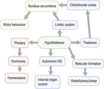
The hypothalamus generates responses to these varied stimuli by, again, both circulatory and neural means (Fig. 16.8). An intimate relationship with the pituitary gland and privileged access to its circulation (portal system) confers the role of ‘orchestrator of the endocrine system’ on the hypothalamus, as it directs hormonal synthesis and release. The neural output of the hypothalamus is directed to widespread regions of the neuraxis. Descending fibres pass to the brainstem and some reach the spinal cord. In this way, connection is made with various brainstem nuclei, including the reticular formation, influencing wakefulness and sleep, and control is exerted over preganglionic sympathetic and parasympathetic neurones of the autonomic nervous system. Ascending connections pass to the limbic system, both directly and, via the thalamus, to the orbital frontal cerebral cortex. The hypothalamus can initiate appropriate motor behavioural repertoires of an instinctive kind through its connections with the limbic system and limbic part of the corpus striatum (the nucleus accumbens). Furthermore, it can influence, or even override, more complex adaptive behaviour because of its close links with the limbic system and the orbital frontal association cortex.
Hypothalamic nuclei
The hypothalamus consists of many nuclear divisions, only some of which will be described here (Fig. 16.1). The region lying medial and ventral to the structures of the subthalamus is known as the lateral hypothalamus. It is traversed longitudinally by many fibres, including the medial forebrain bundle. The lateral hypothalamic area is important in the control of food and water intake and is, in part, equivalent to the physiologically defined feeding centre. Lateral hypothalamic lesions cause aphagia and adipsia.
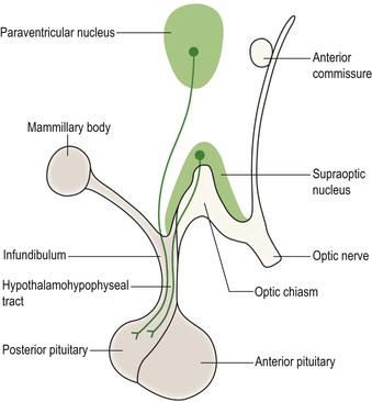
The medial region of the hypothalamus contains various nuclei, only some of which have well-defined functions. Anteriorly lie the supraoptic, paraventricular and suprachiasmatic nuclei. The supraoptic and paraventricular nuclei both produce systemically acting hormones, which are released from the posterior pituitary into the general circulation. The supraoptic nucleus produces vasopressin (antidiuretic hormone), which increases water reabsorption by the kidney. The paraventricular nucleus synthesises oxytocin. In the female, activation of the paraventricular nucleus, and release of hormone, is induced by suckling. This stimulates milk production by the mammary gland and causes contraction of uterine muscle.
The axons of cells in the supraoptic and paraventricular nuclei pass to the neurohypophysis in the hypothalamohypophyseal tract (Fig. 16.2). The neuroendocrine products are transported in this tract to the neurohypophysis, where they are released into the capillary bed and, thus, reach the general circulation.
The supraoptic nucleus contains osmosensitive neurones that are activated by changes in the osmolality of circulating blood. An increase in osmolality causes release of vasopressin. This acts upon the kidney tubules to increase water reabsorption, thus maintaining water homeostasis.
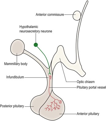
The hypothalamus also synthesises releasing factors and release-inhibiting factors, which control the release of hormones by the adenohypophysis. The adenohypophysis produces adrenocorticotropic hormone (ACTH), luteinising hormone (LH), follicle-stimulating hormone (FSH), thyroid-stimulating hormone (TSH), growth hormone and prolactin, which are released into the general circulation. The factors that control them are released from the terminals of hypothalamic neurones into the capillary bed of the pituitary portal system (Fig. 16.3). These vessels, which are intrinsic to the hypophyseal stalk, convey the released agents to the adenohypophysis, where they act upon the hormone-secreting cells. Within this system, the neurotransmitter dopamine is synthesised by neurones of the hypothalamic arcuate nucleus and is released within the neurohypophysis by axons travelling in the hypothalamohypophyseal tract (Fig. 9.14). Dopamine acts to inhibit the release of prolactin by the adenohypophysis. The synthesis of hypothalamic releasing factors is under feedback regulation by hormones produced by target organs.
 Tumours of the hypothalamus and pituitary gland
Tumours and other diseases of the hypothalamus and associated pituitary gland lead to under- or overproduction of circulating hormones. These, in turn, produce disorders of growth (dwarfism, gigantism and acromegaly), sexual function (precocious puberty, hypogonadism), body water control (diabetes insipidus and pathological drinking), eating (obesity and bulimia) and adrenal cortical control (Cushing’s disease and adrenal insufficiency). Since the pituitary gland is closely adjacent to the optic chiasm, tumours of the gland (pituitary adenomas) may also lead to bitemporal visual field loss.
Related posts:


Stay updated, free articles. Join our Telegram channel

Full access? Get Clinical Tree


