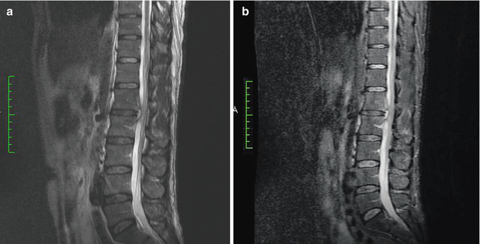
Fig. 14.1
(a) Sagittal STIR FAST image of a male patient showing involvement of two segments. (b) Sagittal T2-weighted image of the same patient. Note the different stages of the disease for each segment

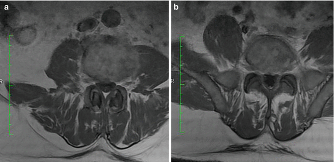
Fig. 14.2
(a) Contrast-enhanced MRI images of the same patient with same level, axial L3–L4. (b) Contrast-enhanced MRI images of the same patient with same level, axial L5–S1

Fig. 14.3
(a) A patient with spinal brucellosis, note the posterior beaklike protrusion, sagittal T2-weighted image, L1–L2. (b) STIR sagittal-weighted image of the same level of the same patient on (a)

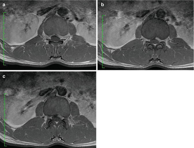
Fig. 14.4
Axial contrast-enhanced images of L1–L2 level with three different axial levels, respectively, lower end plate of L1 (a), mid-level between L1–L2 disc (b), and upper end plate of L2 (c)
14.3.4 Scintigraphy
Radionuclide bone scintigraphy is also an important diagnostic study for detecting spinal infections including brucellosis. The authors who first mentioned the role and importance of nuclear medicine were Bahar and Madkour [22, 23]. Scintigrams may show focal or diffuse increased uptake in the vertebral bodies. The entire vertebral body may be involved, or there may be the caries sign (focal and marked uptake at the intersection region of the upper and lateral margins of the vertebra). Scintigraphy may show involvement of focal long bones, costovertebral joints, and costochondral junction. Sacro-ileitis may also be present and can be seen on radionuclide imaging. Nevertheless, MRI is the choice imaging for spinal brucella.
As mentioned by a recent article and investigation [24], positron emission tomography (PET) can show lesions which are and may not be visualized on MRI. PET/CT combination with metabolites such as fluorine-18 fluoro-deoxy-D-glucose may reveal additional lesions of the spine and paravertebral tissues too.
14.4 Treatment
The first step in the treatment of spinal brucellosis is medication which includes antibiotics effective in intracellular organisms of the brucella genus. As the infection is intracellular, a long medication period is required and close follow-up is needed. One important issue which should be taken into consideration is a sudden neurologic deterioration, in case medical treatment with antibiotics is the initial regimen of therapy. Surgery may be needed in case of neurologic deterioration. In such a case, anterior, posterior, or combined surgeries may be performed.
Involvement of the cervical column is infrequent compared to the lumbar or thoracic region but is more susceptible to severe complications. Epidural mass and spinal cord compression may cause serious neurologic deficits. The anterior surgical approach is the first choice in this region.
Conclusion
The brucella organism can affect all the nervous system regardless of location with manifestations of spinal or central nervous involvement all together named as neurobrucellosis. Laboratory tests and imaging modalities such as MRI, PET, and scintigraphy are used to diagnose this entity.
Although brucella is a systemic infection, local manifestation such as brucellar involvement of the spinal column is a treatable disease and has a good outcome in most cases. Usually, it may cause different complications in the nervous system such as meningitis, granuloma and abscess development, and spondylitis depending on its location, and sometimes all of these manifestations can be seen in one case [25].
Treatment of the infection is mainly conservative with medications unless it has formed an abscess or granuloma. The medications should include drugs acting on intracellular organisms. If antibiotic drugs are administered, blood serum agglutinin levels can be used for the progression of the treatment. Surgery should be kept on hand as an option in case of failure of medical treatment and especially in case of abscesses and neurologic deterioration. In case of surgery, not the only one but the most critical prognostic factor in recovery from the infection is the preoperative neurologic condition.
As a standard manner of diagnostic algorithm, a stepwise imaging modality of choice could be done for the diagnosis of spinal brucellosis, starting from plain X-rays and going through CT and MRI. Plain X-ray is a cheap and easy to access technique for imaging spinal brucellosis, but the overmentioned signs for the disease has a disadvantage; as details may be easily passed over. But along with laboratory tests and with a careful eye, this easy and cheap imaging modality can lead to the diagnosis of spinal brucellosis.
CT, the more precise imaging modality than plain X-ray, comes as the second stage, but recently MRI is almost always the choice as the first step of imaging for spinal brucellosis and all other manifestations of the disease. MRI reveals an early diagnosis of vertebral involvement of spinal brucellosis and has the advantage of avoidance of X-rays of CT. With enhancement of contrast media, it provides a precise diagnosis. Recently additional nuclear medicine imaging techniques such as scintigraphy may help in the diagnosis of the disease earlier than MRI, especially that PET has this advantage. But as MRI has widespread accessibility and advantage of protection from ionizing radiation, it can be used as the diagnostic study of choice for spinal brucellosis.
Stay updated, free articles. Join our Telegram channel

Full access? Get Clinical Tree


