(1)

(2)
Data Analysis
Data are presented as the mean ± standard error of the mean (SEM). Data were analyzed using one-way analysis of variance (ANOVA) with Tukey post-hoc test. A p < 0.05 was considered statistically significant.
Results
Ipsilateral Hemisphere Volume
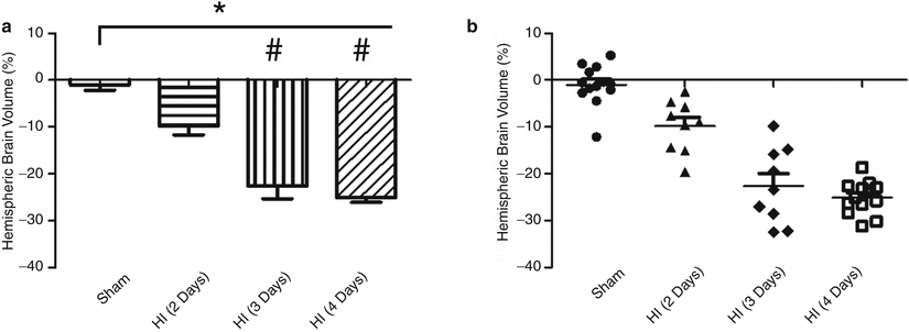
Animals subjected to HI sacrificed at 2, 3, or 4 days after HI had significantly lower ipsilateral brain hemisphere volumes compared with sham animals (sham: −1.0 ± 1.20 %, n = 13) (p < 0.01 vs. sham for HI (2 days), HI (d Days), and HI (d Days)). Ipsilateral hemisphere volumes for the HI animals sacrificed at 2 days (HI (2 days): −9.8 ± 1.89 %, n = 9) were significantly higher than those of animals sacrificed at either 3 days (HI (3 days): −22.6 ± 2.70 %, n = 9) or 4 days (HI (d Days): −25.1 ± 0.97 %, n = 13) (p < 0.01 for HI (3 days) vs. HI (2 days) and p < 0.01 for HI (4 days) vs. HI (2 days)). No statistical difference in hemispheric brain volume was observed between animals sacrificed at 3 days after HI compared with that of animals sacrificed at 4 days post-HI (Fig. 1).


Fig. 1
Hemispheric Brain Volume after HI. Ipsilateral hemisphere brain volume (%) for HI animals is less than that of sham for all HI groups (p < 0.01 for HI (2 days) vs. sham, p < 0.01 for HI (3 days) vs. sham, p < 0.01 for HI (4 days) vs. sham). The hemisphere brain volume for HI animals sacrificed at 3 and 4 days were indistinguishable (p > 0.05) but both of these groups were significantly lower than that of 2 days post-HI (p < 0.01 for HI (3 days) vs. HI (2 days), and p < 0.01 for HI (4 days) vs. HI (2 days)). * p < 0.01 vs. sham, # p < 0.01 vs. HI (2 days). n = 9–13/group. The same data shown as bar graphs (a) and as scatter plots (b)
Infarct Volume
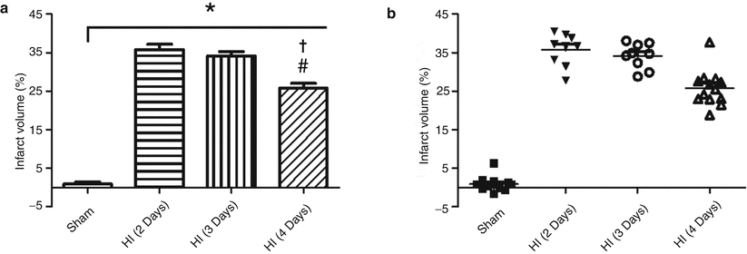
Animals sacrificed at 2, 3, and 4 days after HI had significantly higher infarct volumes than those of sham animals (sham: 0.9 ± 0.52 %, n = 13) (p < 0.001 vs. sham for HI (48 h), HI (72 h), and HI (96 h)). The infarct volumes for HI animals sacrificed 2 days (HI (2 days): 35.8 ± 1.39 %, n = 9) and 3 days (HI (3 days): 34.2 ± 1.09 %, n = 9) were not significantly different from one another (p > 0.05 HI (2 days) vs. HI (3 days)). However, 4 days post-HI, the infarct volume (HI (4days): 25.8 ± 1.28 %, n = 13) was significantly lower than that of HI animals sacrificed at either 2 days or 3 days post-HI (p < 0.01 for HI (4 days) vs. HI (2 days) and p < 0.01 for HI (4 days) vs. HI (3 days)) (Fig. 2).


Fig. 2
Infarct volume after HI. Infarct volume (%) for HI animals is greater than that of sham for all HI groups (p < 0.01 for HI (2 days) vs. sham, p < 0.01 for HI (3 days) vs. sham, p < 0.01 for HI (4 days) vs. sham). The infarct volume for HI animals sacrificed at 2 and 3 days were indistinguishable (p > 0.05) but both of these groups were significantly higher than that of 4 days post-HI (p < 0.01 for HI (4 days) vs. HI (2 days), and p < 0.01 for HI (4 days) vs. HI (3 days)). * p < 0.01 vs. sham, # p < 0.01 vs. HI (2 days), † p < 0.01 vs. HI (3 days). n = 9–13/group. The same data shown as bar graphs (a) and as scatter plots (b)
Discussion
Herein, we investigated the effect of post-HI sacrifice time on brain volume and infarction volume using a neonatal rat pup model of HI. To our knowledge, this is the first time the role of sacrifice time after HI has been investigated for both brain hemisphere volume and infarct volume.
Effect of Sacrifice Time on Ipsilateral Hemisphere Brain Volume after HI
Volume of the ipsilateral hemisphere is expected to decrease in the acute phase following HI [1, 9, 13], and this study supports the literature. The most plausible explanation for the smaller brain volume of the ipsilateral, injured hemisphere is that the brains of neonates are rapidly developing, gaining mass with each passing day. Any cerebral injury that causes tissue death will likely cause the ipsilateral brain volume to be smaller than the healthy, contralateral hemisphere. Additionally, tissue damage may retard growth in the ipsilateral hemisphere because of conflicting mechanisms of the injury (deleterious molecular mechanisms causing inflammation and/or cell death, and repair mechanisms) and developmental mechanisms.
However, the changes in brain volume over the days after HI have not been investigated in either patients or animal models. Herein we observed that the ipsilateral hemisphere brain volume decreases from day 2 to day 3 post-HI and seems to level at the third day following HI. While the reason for the decrease from 2 to 3 days after HI is unclear, it may include tissue growth and repair mechanism, but also may be a limitation of the current algorithm used for calculating ipsilateral hemisphere brain volume.
Effect of Sacrifice Time on Infarct Volume after HI
The infarction after HI includes a penumbra and a core in which the penumbra contains injured but salvageable tissue. The exact time in which cells transition from salvageable to irreversibly damaged is variable, but likely occurs over the first 5 days after HI [8]. Although the change from penumbra to core infarct may begin as early as a few hours after insult [2], Ghosh et al. [8] found that the overall infarct size is a maximum 2 days after HI and then decreases. One caveat of the study by Ghosh et al. is that the equation used to calculate infarct volume does not correct for swelling/volume changes. Regardless, the findings in this study agree with those of Ghosh et al. The changes in infarct volume are likely to the result of a combination of brain growth and repair mechanisms.
Stay updated, free articles. Join our Telegram channel

Full access? Get Clinical Tree


