For all patients
Noncontrast brain CT or brain MRI (followed by angiography)
Blood glucose
Oxygen saturation
Blood pressure and pulse rate
Serum electrolyte/renal function test
Hepatic function test
Complete blood count (especially platelet count)
Cardiac ischemia markers
Electrocardiogram
Coagulation tests including prothrombin time, activated partial thromboplastin time, and D-dimers
Chest radiography
For selected patients
Toxicology screening
Blood alcohol level
Pregnancy test
Arterial blood gas tests
Lumbar puncture
Electroencephalogram

Fig. 3.1
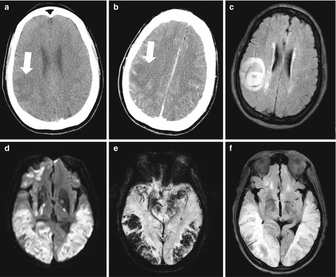
Representative cases of stroke mimics. The first case (a–c) is a 65-year-old woman who complained of sudden onset left-side weakness and hypesthesia. Initial pre- and post-gadolinium (a, b) brain CT showed low-attenuated lesion involving right postcentral gyrus without enhancement (white arrow). Since initial symptom onset was within 3 h, she was treated by intravenous thrombolysis, but weakness progressed thereafter. Subsequent brain MR imaging (MRI) with T2-based fluid-attenuated inversion recovery protocol showed edematous mass lesion with heterogeneous signal intensity and fluid-fluid level. Further evaluation with serial brain MRI and biopsy revealed brain abscess as a final diagnosis. The second case (d–f) is a 62-year-old woman who was found to be in a comatose state at home. Initial neurological examination revealed semicomatose mental status and left-side dominant weakness with extensor Babinski reflex. Brain MR imaging showed diffuse high-signal intensity from diffusion-weighted image and low-signal intensity from susceptibility-weighted image mainly involving both parietal and occipital cortices. Admission serum glucose level and osmolality at admission were significantly elevated, and she was diagnosed as hyperosmolar hyperglycemic coma
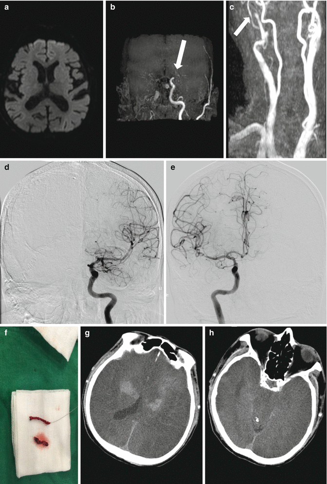
Noncontrast brain CT has been recommended as an initial imaging modality to rule out hemorrhagic stroke and to initiate intravenous thrombolysis. Recent advance of endovascular thrombectomy among acute ischemic stroke patients confirmed therapeutic efficacy and safety from multiple large randomized clinical trials; therefore, prompt evaluation of cerebral arteries is getting more critical among the patients with suspected major intracranial arterial occlusion (Fig. 3.2). So far, there exists a wide variation of brain imaging protocols for acute stroke patients among stroke centers. Further research needs to be performed to determine appropriate imaging protocol beyond noncontrast brain CT to select optimal additional imaging modalities among CT angiography, brain MRI, or perfusion imaging and also to prioritize among additional imaging studies and intravenous thrombolysis.


Fig. 3.2
A case of missed stroke diagnosis. An 85-year-old man with lost consciousness was transferred from general hospital. Neurologist was delayed 4 h after symptom onset because primary assessment was syncope, but recovery of mentality was delayed. Initial neurological examination revealed semicomatose mental status with decorticated posture and bilateral extensor Babinski reflex. Brain MR imaging with diffusion-weighted image showed slight increased signal intensity involving both cerebral hemispheres (a). MR angiography revealed the occlusion of left middle cerebral artery and right internal carotid artery at petrous segment (b, c). Emergent endovascular thrombectomy successfully recanalized both occlude vessels (d, e) and extracted red thrombi (f), but his mentation was not recovered and followed brain CT revealed massive brain edema and hemorrhagic transformation involving both cerebral hemispheres (g, h). He died 5 days after symptom onset
3.2 Cases with High Risk of Missed Diagnosis of Stroke
Missed stroke diagnosis in emergency setting could result in catastrophic consequence because of delayed reperfusion treatment or missed opportunity for secondary prevention with antithrombotics. The length of stay is increased, and the neurological deficit at discharge and mortality rate is higher among missed stroke victims than those without missed diagnosis. Several retrospective studies show that initial misdiagnosis occurs in up to 20% of stroke patients, without significant differences between emergency department of academic center where neurologist primarily deals with the suspected stroke patient and community hospital where general physician manages stroke suspects [4]. Based on several hospital-based cohort studies, the risk of missed diagnosis is greatest in the following conditions: (1) posterior circulation stroke, especially when initial symptom was isolated vertigo or loss of consciousness, and (2) a patient with young onset age.
Stay updated, free articles. Join our Telegram channel

Full access? Get Clinical Tree


