Chapter 191 Interventional Nonoperative Management of Neck and Back Pain
Pain and disability from spine pathology are usually benign, temporally limited occurrences that may be accompanied by anxiety and hypervigilance on the part of the sufferer. Occasionally, symptoms of pain are caused by tumor, infection, or trauma, requiring aggressive and often morbid interventions. On the other hand, most acute episodes of spinal pain improve substantially within 90 days.1,2 When the complaint of spinal pain is accompanied by minor/mild neurologic deficit and ample evidence of degenerative change, the answer may become confounded by contradictory evidence. In such cases, interventional procedures are increasingly utilized to relieve symptoms, ameliorate disability, and reduce surgical risk.3,4 This chapter presents an overview of available interventional spine procedures, their indications, and the best available evidence for efficacy within a continuum of clinical care.
Conservative Care Progression
When discussing treatment options with patients in acute distress, it is important to keep in mind the first tenet of Western medicine: “first, do no harm.” This is best accomplished by following a progression of conservative care that first presents strategies that provide the least risk with the highest likelihood of success. In the rush to ameliorate pain, experienced physicians occasionally forget that any intervention should have patient function as the primary goal and pain relief as a laudable, but secondary, goal (Fig. 191-1). Moreover, it has been shown that people will accept a degree of chronic pain if they are able to participate in desired activities.5 The decoupling of symptoms and radiologic/anatomic findings can lead similarly well-trained professionals to vastly different opinions. In the end, the patient is best served when the treatment modality with the least risk is applied to relieve a given pathology. Following the “do not harm” tenet, efficacious, timely applied spinal injections may allow enough time for the body to heal and return to function without the need for further invasive procedures.
Overview of Selective Spinal Procedures
As stated previously, selective spine procedures are being used with increasing frequency to manage acute and chronic pain syndromes.3 The implied societal benefit of a greater number of procedures is more robustly measurable functional improvements. To the contrary, literature states that increasing expenditures for interventional and surgical procedures have not translated into improved health status for spinal pain sufferers.6,7 Moreover, disability from spine-related pain is increasing in the United States.8 Overutilization of procedures is a problem that undermines the fundamental doctor-patient relationship as well as the collegial doctor-doctor relationship. Failure to live up to the promise of increased function leads to a rapid erosion of the trust that underlies everything physicians do. Therefore, this chapter follows the principles of Edward Benzel, MD, when discussing and describing indications and evidence for selective spine procedures. These principles enable increasing trust: Wisdom has foremost importance, but should be combined with evidence, and statements of risk/efficacy should be based on what one would do to their own mother, spouse, or child.9
The pain generator, the Holy Grail of clinical and diagnostic medicine, is often illusory in spinal pain patients. Multiple studies by Boden, Wiesel, Mayer, Rainville, and Carragee10–14 have shown the discordance between pain and anatomic or physiologic abnormalities. Given that pain is as much a cognitive as it is a nocioceptive phenomenon, if pain response is the sole outcome measure, the selection bias will always trend toward failure. This chapter, therefore, attempts to utilize evidence that has a functional component (sparse in spine literature) accompanying the usual visual analogue scale (VAS) when discussing the various procedures available to spine surgeons to improve their outcomes.
One must also understand the masquerade, or the concept of pain referral. Often a lumbar issue can masquerade as or coincide with a hip problem.15,16 This concept applies to the neck and shoulder as well as to various nerve entrapment syndromes in the upper or lower extremity.17 Acumen to unlock the masquerade is found in the office and not in the operating suite. Therefore, a skilled interventionist who performs a thorough physical examination can accrue good internally valid, reproducible diagnostic information to keep a surgeon from making a hasty, unwise decision.
The inflammatory basis for pain’s relation to the degenerative changes in spinal structures and diarthrodial joints is an additional concept to critically consider. Although concepts of legal indemnity and political policy lag behind, there is substantial evidence that degenerative changes to intervertebral discs, cartilage, and joints begin with inflammatory changes and not with mechanical stress/injury (cumulative or otherwise). This subtle concept is important for two reasons. First, the radiologic appearance of the structure is less important than its functional range and strength, which may recover through a combination of aggressive treatment of acute-phase inflammation with maintenance of functional range of motion over the same time. Second and more importantly, physician advocacy of inactivity to prevent further injury undermines the ultimate benefit of any proposed intervention. Specifically, patients who have been advised by physicians that activity (not inflammation) causes injury have higher pain scores and trends to disability.18 Physician advice that worsens the cognitive aspect of pain begin a self-fulfilling pain/disability cycle that rarely ends well.
Radicular Nerve Pain and Therapeutic versus Diagnostic Injections
Radiculopathy is a pathologic process affecting a spinal nerve root in which sensory and/or motor symptoms present distant from the pathology. Whether the process is mechanical/compressive, inflammatory, or both remains poorly elucidated. What is clear is that the sodium-channel blocking effects of local amide or ester anesthetics provide relief from pain but do not improve any functional deficits. Due to the pharmacokinetics of existing local anesthetics, this benefit is temporary but can (when applied discreetly and correctly) provide excellent diagnostic information. Synthetic glucocorticoids act to suppress production of inflammatory mediators, stabilize membrane irritability, and enhance macrophage demargination to the site of injury. Due to the complexity of the interactions, there is often a delay before the benefit from locally or remotely administered glucocorticoid is perceived by the patient.19 Depending on the nature of the nerve irritation/injury, the benefit of the glucocorticoid can often be long-lived. A tapering dose of medication that up-regulates systemic circulating glucocorticoid is still in common use for suspected radiculopathy; however, rapid steroid metabolism and intolerable systemic side effects occasionally make systemic use impractical or too short-lived for functional improvement. For this patient, treatment targeted to the pathology is often beneficial.
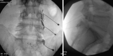
Epidural steroid injection for radiculopathy was first described in the United States in 1960 in the Cleveland Clinic Quarterly and was expanded upon thereafter.20,21 In the years since that time, its efficacy has been enhanced by use of imaging guidance as well as training programs that ensure a physician’s safe delivery of medication closest to the site of pathology. The intent of delivery of glucocorticoid medication to the site of pathology is the deposition of fat-soluble steroid in a nearby plane of epidural fat. Glucocorticoid steroids will release from this adipose tissue in a prolonged fashion to reduce pain by down-regulating pro-anti-inflammatory, acute-phase chemical mediators from inflamed/reactive tissue. Broadly, the three ways of accessing the epidural fat are through the sacral hiatus (caudal injection), between the bony lamina and traversing the ligamentum flavum (interlaminar injection), and through the neural foramina near the bifurcation of the nerve root from the spinal cord or dural sac (transforaminal injection). Each method has its utility, with research showing a trend toward greater efficacy, fewer systemic effects, and increased cost utility when the injection delivers medicine closest to the pathologic process responsible for pain and functional inhibition. In general, all of these types of injections should be assisted by imaging guidance. The studied rate of misapplication of medication with resultant complications varies from 22% to 69%. Therefore, an interventionist’s failure to use imaging guidance should be a red flag to the referring physician (Fig. 191-2).
Caudal Epidural Steroid Injection
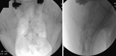
The starting needle placement for a caudal injection can be guided by palpable anatomic landmarks or by imaging. With this in mind, there is a greater than 25% rate of needle-tip misadventure without imaging guidance.22 The procedure is fairly simple, with a blunt-tipped needle directed through the sacral hiatus assisted by a simple two-picture confirmation in anteroposterior and lateral views. The needle is advanced along the dorsal bony elements of the sacrum to approximately the S3-4 space. The thecal sac typically ends around S2, but may extend as low as S4. Nonionic contrast material is used to confirm that the needle is placed within the epidural space and outside the thecal sac. Violation of the dura mater results in a spinal headache. Injection of a steroid and anesthetic solution into the caudal epidural space may result in urine retention. Additionally, delivery of the medicine through the caudal approach is often remote from the location of pathology. Finally, the concentration of corticosteroid is significantly less well distributed in the sacral epidural fat, with a concentration decrement of tenfold or more of the injectate reaching the site of pathology (Fig. 191-3).
Interlaminar Epidural Steroid Injection
The steps for getting good results from interlaminar epidural steroid injections are similar to those for other techniques. First, the physician should plan the procedure by taking a history, performing a thorough physical examination, and correlating that information to appropriate imaging. If the predominant complaint is axial back pain, the chances of a medium or long-term, nonplacebo, beneficial result from depot of steroid in the epidural space is low. Alternatively, when the pain presentation is predominantly in the limb and subjective complaints correlate with discreet pathology on radiology, the chance of injection benefit and avoiding surgery is high.23
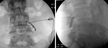
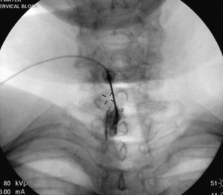
Once planning is complete, the procedure is straightforward. Fluoroscopic guidance facilitates the localization of the desired level in the lumbar, thoracic, or cervical spine. The skin overlying the interspace between the spinous processes is anesthetized, and most commonly a blunt-tipped needle is passed through the skin and directed midline. The needle is advanced using a combination of direct, fluoroscopic visualization and “loss of resistance” technique with a syringe containing water or air. Skillful completion of these techniques is important in reducing the incidence of spinal headache by dural puncture. A sound understanding of anatomy is vital for this procedure, because the spinous processes are oriented differently in the cervical, thoracic, and lumbar spinal divisions. Additionally, the thickness of ligaments, depth of lateral recesses, and location of vascular anatomy changes over the course of the spine. To obtain an efficacious result, medication needs to be placed in the epidural space only. To ensure proper placement, it is strongly recommended that a form of nonionic contrast be used to verify placement once loss of resistance is encountered. Frequently, even in the most experienced hands, a false loss of resistance may be encountered between the interspinous ligament and the ligamentum flavum or, more problematically, tissue may become lodged in the bevel of the needle, resulting in a failure to perceive loss of resistance. This failure often results in dural puncture and in the contrast flow pattern appearing identical to a myelogram. Finally, knowledge of anatomy allows one to understand that the epidural space is not uniform in shape but is pyramidal in cross section, meaning that if the needle tip is directed away from midline, the likelihood of dural puncture increases in proportion to the decreased epidural space laterally. McGoldrick expanded on the cryomicrotome dissection work of Hogan to show that failure of ligamentum fusion in the dorsal midline occurs in the cervical and high thoracic space in 51% to 74% of dissections.24,25 Additionally, controversy persists about whether a “fat pad” exists dorsally above the dura mater at a level above C7.24 The lack of landmarks for traditional loss of resistance technique make dural puncture in the cervical and high thoracic spine more likely. The consequence of dural puncture with intrathecal infusion of anesthetic can range from slow-onset respiratory depression (~30 minutes) to seizure to cardiovascular collapse.26
Finally, Cluff et al. noted a disturbing trend: that only 39% of academic institutions used fluoroscopy for epidural injections, whereas 74% of private practices used fluoroscopy.27 In light of the compelling data that only 26% of blind attempts (without fluoroscopic guidance) by experienced anesthesiologists placed medication at the site of pathology, this trend may account for the jaundiced view held by many referrers with regard to the efficacy of injections.28 This jaundiced view is seen at its most extreme in the oft-cited study by Valat that, in attempting to demonstrate the efficacy of epidural steroid injection versus placebo merely showed that blind epidural steroid injection is no better than placebo even when delivered three times.29 In conclusion, the prosaic use of the interlaminar technique has bred contempt that can only be improved if referring physicians demand excellence; one may consider the oft-quoted Edward Benzel aphorism: “A fool with a tool is still a fool.”
Transforaminal Epidural Steroid Injection
The transforaminal epidural steroid injection technique (Fig. 191-4) has been used with increasing favor owing to beliefs around the selectivity, proximity (to the pathology), and longevity of benefit with this technique. These assumptions have proven difficult to substantiate in the literature. Riew et al. published two very persuasive studies that were shrewdly designed to eliminate the winner’s bias seen in many studies examining injection versus surgery.30 The authors took all patients who were scheduled for surgery and randomized them to receive a transforaminal injection of either anesthetic alone or anesthetic plus steroid. Of those receiving steroid via transforaminal delivery, 71% elected to not have surgery at end points ranging from 13 to 27 months. This was a statistically significant difference compared to the anesthetic-alone group, in which only 33% elected not to have surgery by the end point (p < .004). This finding held at 5-year analysis, with 3 of 4 patients requiring late surgery (between 27 and 73 months) with progression of symptomatic spinal stenosis.23
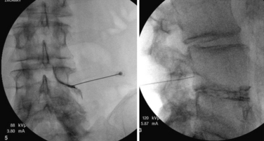
FIGURE 191-4 Right L4 transforaminal epidural steroid injection: anteroposterior (left) and lateral (right).
Recently, there has been a proliferation of expert opinion about the risks associated with cervical epidural steroid injections in general and cervical transforaminal injections specifically. The main concerns originate from case reports of catastrophic complications, including death, persistent vegetative state, stroke, spinal cord infarction, and high anesthesia-related cardiorespiratory collapse. For the most part, the culprit is injection of medication into the arterial circulation supplying the brain or spinal cord. Several safeguards exist and should be known by a fellowship-trained interventionalist—including use of contrast enhancement to confirm needle-tip placement, use of “live” fluoroscopic visualization to confirm arterial uptake, use of test-dose anesthetic medication, and use of digital subtraction technology. Each of these recommendations has its benefits; however, the most convincing evidence to recommend modifying current practice comes from an animal study that demonstratied zero infarction rate with administration of nonparticulate steroid in two different formulations; 100% of the particulate steroid group suffered neurologic damage that correlated with hypoxic and ischemic damage.31
In conclusion, epidural steroid injections show benefit when performed for the correct reason (radicular, not axial, pain) and when medicine is delivered to the correct location (using fluoroscopic guidance and contrast confirmation). These injections have been shown to help reduce surgical rate, allow for earlier return to function, and save $12,666 per responder (in 1999 dollars).30,32,33 Therefore, referring physicians should insist on the highest clinician acumen and the highest-quality training for proper delivery of medication to the site of pathology.
Joint-Mediated Pain
The joints of the spine include a three-part joint that combines the disc and a matched pair of dorsal joints at each spinal segment colloquially called the facet joints. Additionally, the cervical spine has accessory joints that aid with shear forces; these are named after the anatomist Luschka, or are commonly called the uncovertebral joints. A separate joint comprises the skull, first vertebra, dens, and second vertebra and allows for complex head movement in relation to the neck and the rest of the body. Finally, a pair of complex joints that transmit/disperse rotational force applied through the pelvis are called the sacroiliac joints. All of these joints are diarthrodial joints of the spine, and each one can produce pain. Briefly we discuss the role of injection procedures in diagnosis and treatment of pain syndromes involving the various spinal joints.
Facet Joints of Cervical, Thoracic, and Lumbar Spine
The facet joints are paired diarthrodial joints on the dorsal aspect of the vertebral bodies. These joints provide stability and are a key component to ensuring accommodation of a neutral zone in the three-joint complex model of the vertebral motion segment. As early as 1911, Goldthwait postulated that the facet joints, specifically the zygapophyseal joints (Z joints) of the lumbar spine, could be a potential pain generator in the spine.34 Anatomic studies have verified the presence of nociceptive structures and mechanoreceptors in the capsule and synovial folds of the facet joints.35–37 Additionally, Mooney and Hirsch et al. identified the zygapophyseal joint as a source of pseudoradicular pain in both a control hypertonic saline group and in study patients.38–40 And why wouldn’t high-stress joint complexes be a potential source of pain? It stands to reason that like the knee, the shoulder, or any diarthrodial joint, the facet joint is subject to the ravages of time, inflammation, and apoptosis, giving it the ability to act as a pain generator in the spine.
Statistical methodology varies, but epidemiologic studies note that facet-mediated pain may account for between 4%41 and 45%42 of chronic spine pain.43 The large variance in these statistics may be due to study inclusion criteria and the lack of adherence to exacting standards. Although proof for the existence of facet joint–mediated pain is debated, the ubiquity of facet joint arthropathy is undeniable. Cadaveric studies have shown facet joint arthropathy to be present in 100% of cadaveric spines over 60 years old.44 Although clues to facet-mediated pain may be obtained from a thorough history, physical examination findings, and imaging, no pathognomonic test/maneuver exists. Facet joint pain is currently diagnosed by a preponderance of evidence because we lack a single test for facet pain that combines high sensitivity and specificity. The lack of any test with a high positive prognostic value is likely due to known compensatory mechanisms that are associated with joint pain/inflammation, such as muscle spasm, altered movement mechanics, and pain referral patterns. Facet joints present more of the prerequisites to be a pain generator than pain associated with the more amorphous construct of discogenic pain. Facet joints have a robust nerve supply and are capable of causing pain similar to that seen in peripheral joints, as in overuse injuries elsewhere in the body. Articular joints possess a known pattern of disease/injury susceptibility, and their pain can be ameliorated using well-described diagnostic techniques with acceptable reliability and validity (Fig. 191-5).45
Unfortunately, today there is no “gold standard” criterion to confirm with high positive predictive value the presence of facet-mediated pain. The accepted standard combines clinical judgment with a diagnostic or therapeutic facet joint injection. Early studies performed by Hirsch et al. and Mooney not only supported the existence of facet joint pain but also seemed to show improvement when injections of anesthetic or glucocorticoids were combined with exercise. Similar results were seen with provocative injections to the medial branch nerves that supply the facet joints.46 Though prone to operator error, these studies show that wise use of facet joint injections can serve dual diagnostic and therapeutic goals in returning patients to function. Currently two techniques are employed to block pain from facet joints: intra-articular injection of the facet joint capsule and blockade of the medial branch nerves transmitting pain signals from the joint to the spinal cord/brain. Both techniques have been shown to possess variable, operator-dependent accuracy/efficacy when combined with appropriate clinical judgment.47,48
The early standard for the diagnosis of facet joint pain was to seek mitigation of pain with a single facet joint injection. However, this methodology produced an unacceptably high false-positive rate of up to 38%,49 meaning results showed poor prognostic value. Coupled with a known/accepted positive placebo response rate of up to 32% with many interventional procedures, this result significantly reduced the validity of this treatment protocol.50 It has therefore been advocated that a reliable control be employed in the form of the double-block protocol, in which anesthetics of varying durations of effect are administered on subsequent injections and the patient maintains two accurate postinjection pain diaries. If the duration of pain relief is concordant with the half-life of the diagnostic anesthetic, the injection is considered diagnostic for facet pain. In addition, many of the best-performed postoperative studies advocate the use of a cut-off score of 75%, 80%,51 or even 100%52 pain relief to indicate a positive response, as opposed to the earlier practices of 50% pain relief.51–54 Furthermore, the importance of outcome measures cannot be stressed enough. As was alluded to earlier in this chapter, the focus of diagnostic and therapeutic injections should be functional improvement rather than mitigation on VAS scores. Therefore, evaluation with validated functional measures such as the Pain Disability Questionaire (PDQ), Pain Disability Index (PDI), Euro-Qual, SF-18, or even the old standard Oswestry Disability Index should be utilized when interpreting the effectiveness of any of the interventional procedures discussed in this chapter.
Much debate has arisen regarding the selectivity of intra-articular injection versus medial branch blocks. Cadaveric studies by Dreyfuss et al. suggest that volume control is important in limiting spread of local anesthetic to nearby structures to avoid anesthetizing other nociceptive structures and providing a false-positive result by exerting pain relief and functional benefit to structures other than the facet joint.53 This research has led many clinicians to follow the “less is more” axiom and to use less than 1 mL of anesthetic for their diagnostic blockade. This small aliquot of anesthetic medication seems to have the best postprocedure positive predictive value, correlating to prospective successful outcomes.51
Understanding the myriad of variables that complicate all clinical research, many functional outcome studies have been performed with questionable enrollment criteria. Meta-analyses utilizing stringent compliance with Agency for Healthcare Research and Quality (AHRQ) and Quality Assessment of Diagnostic Accuracy Studies (QUADAS) criteria for evaluation of diagnostic tests suggest that there is strong evidence that controlled diagnostic facet joint blocks establish a diagnosis of facet joint pain in chronic cervical and lumbar spinal pain and moderate evidence for diagnosing thoracic facet joint pain.55–57 A review by Boswell et al. and another by van Tulder have supported the diagnostic efficacy of facet joint blockade.58,59 Some reviews still conclude that the multifactorial nature of degenerative joint-associated pain means that there is insufficient evidence to support the use of facet joint injections to diagnose or treat facet joint pain.7,60
As with epidural injections, it is important that the reader note that the use of fluoroscopy or CT imaging guidance is critical to success when performing any diagnostic spinal procedure. Significant care should be taken when selecting an interventionist to perform diagnostic injections on patients who you may ultimately be taking to surgery. These procedures cannot be performed with any degree of accuracy without the use of imaging to verify appropriate needle placement and ensure patient safety. Most interventional societies also advocate the additional use of nonionic contrast to verify placement of injectate. This has additional benefit if the interventionalist and/or the referring physician need to review the procedure at a later date. In preparation for an injection, prior review of available advanced imaging studies (CT or MRI) is important to understand the three-dimensional anatomy of the target joints. This is especially true with intra-articular facet joint injections because there can be considerable variability in joint morphology among individuals. The C arm is usually aligned in a slightly oblique fashion to afford the physician access to a portion of the facet joint capsule. The facet joint capsule can be accessed at various locations along the joint line or at the superior or inferior recess. The amount of subcutaneous anesthetic required to keep a particular patient comfortable varies from practitioner to practitioner and from patient to patient. Overinjection of large amounts of anesthetic into the subcutaneous structures and paraspinal musculature has been criticized as a likely contributor to the false-positive rates seen in some studies.61 Yet another reason to follow the “less is more” dictum, with use of a 25G needle smaller amounts of subcutaneous anesthetic are required. The needle is advanced under fluoroscopic guidance and is often felt piercing the joint and capsule. Confirmation of intra-articular needle placement is made with a small amount of contrast material. The resulting arthrogram will typically reveal retained contrast material in the superior and inferior capsular recesses of the facet joint. At times, a well-demarcated plane of contrast will be seen between the two articular surfaces. Fluoroscopic confirmation is best made in at least two planes, preferably 90 degrees from each other. As stated previously, retention of fluoroscopic images can be critical for future evaluation of the efficacy of the injection and for quality control by the referring surgeon. Once intra-articular flow of contrast is confirmed, a small amount of anesthetic or a solution of anesthetic and long-acting glucocorticoid is injected into the joint before the needle is removed. Typically the amount of this solution is less than 1.25 mL.
An additional confounding issue surrounds the routine use of moderate or deep sedation by anesthesiologists during interventional procedures; this may have unintended consequences and can worsen the predictive value of these procedures. Manchikanti et al. studied the effects of IV midazolam and/or fentanyl during cervical and lumbar facet joint injections and showed that with strict criteria the effects of sedation may be minimized to a 10% false-positive rate.61
We do not advocate consideration of medial branch nerve radiofrequency ablation (or rhizotomy) unless suspicion of facet joint–mediated pain is supported by clear clinical suspicion, imaging evidence, and two successful diagnostic blocks. This literature-supported opinion has been brought into question by a recent paper by Cohen et al., who contend that the cost of blocks to increase the positive predictive value exceeds the cost utility from pain relief and increased function seen when one proceeds straight to radiofrequency rhizotomy of medical branches.62 First described by Shealy, this procedure denervates the neural pathways (medial branch nerves) that supply the afferent pain information from the facet joint to the central nervous system.63 This procedure has been shown to offer longer relief than intra-articular facet joint injections and medial branch blocks.51 The consistent locations of the medial branch nerves in the lumbar and cervical spine, reported by many anatomists, make these excellent locations for dorsal rami radiofrequency rhizotomy. However, one must recall the redundant nature of the medial branch innervation of the facet joints. Namely, each joint receives innervation from two different dorsal rami levels. For example, the L4-5 facet joint receives its innervation from the L3 medial branch from above and the L4 medial branch at that level. The innervation of the cervical facet joints is somewhat different. The cervical facet joint is innervated by the medial branch nerve of that level and the level below. The reason for this variance lies in the presence of the C8 nerve, which innervates the T1-2 facet joint in conjunction with the T1 medial branch. This consistency of location explains the favorable success rates with cervical and lumbar medial branch blocks and radiofrequency procedures. On the other hand, the location of the thoracic medial branch nerves is known to be quite variable along the length of the thoracic transverse process, which makes effective lesioning of these nerves quite challenging technically. There is ongoing debate as to the most effective techniques to effectively denervate the thoracic facet joints. Directing the radiofrequency rhizotomy probe to its target is performed in much the same manner as in the medial branch block procedure. The major difference is considerations of cross-sectional area in placing a maximal amount of the uninsulated tip of the special insulated radiofrequency needle in the area where the nerve resides. Care is taken to avoid adjacent structures such as nerve roots and deep muscles that were partially lesioned and can cause a new and persistent pain.64 Again, placement of these needles is performed using fluoroscopy and radiopaque contrast. A lesion is created using either a continuous or pulsed radiofrequency apparatus at 80 to 90 degrees for 80 to 90 seconds. Some practitioners repeat this procedure with slight adjustment to the location of the active tip to create several lesions along the same nerve.
A review by Slipman et al. reports moderately strong evidence (level III) in accordance with the guidelines described by the AHCPR that radiofrequency ablation of the medial branch nerves is an effective treatment for facet joint pain.65 Other reviews have supported the presence of only moderately weak evidence or conflicting evidence regarding the efficacy of a radiofrequency rhizotomy of medial branch nerves for facet-mediated pain.66–68 The goal again is increased function and avoidance of the surgical alternative, which is often a fusion. Many continue to argue that the studies reviewed showed poor efficacy due to the authors’ failure to employ the double-block criteria that are advocated to identify patients with facet joint–related pain. Therefore, many studies that refute the efficacy of radiofrequency rhizotomy of the medial branch nerves for facet joint pain may have not been treating facet joint pain. In a study by Dreyfuss et al. that employed strict standards and double-block techniques, 60% of patients experienced at least a 90% pain reduction, and 87% of patients experienced at least a 60% reduction in pain after a radiofrequency ablation procedure for low back pain, lasting at 12-month follow-up.51 Patient safety and efficacy of procedure should remain stronger considerations than cost despite ongoing pressure from insurers. It is the authors’ opinion that double-block protocol to determine the injection location and pain generator remains the standard of care.
Despite the continuing debate regarding the efficacy of these procedures, there is a consensus that facet joint injections, medial branch blocks, and radiofrequency ablation are safe, reliable, and minimally invasive options to diagnose and treat patients with facet joint pain (Fig. 191-6).
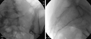
FIGURE 191-6 Sacroiliac joint injection with sacroiliac arthrogram illustrating intra-articular placement.
Related posts:
 Definition and Assessment of Dysfunctional Segmental Motion
Pathophysiology of Cervical Myelopathy: Biomechanics and Deformative Stress
Combined Ventral-Dorsal Surgery
Bone Void Fillers: Bone and Bone Substitutes
Medical Management of Neck and Low Back Pain
Posterior and Transforaminal Lumbar Interbody Fusion
Definition and Assessment of Dysfunctional Segmental Motion
Pathophysiology of Cervical Myelopathy: Biomechanics and Deformative Stress
Combined Ventral-Dorsal Surgery
Bone Void Fillers: Bone and Bone Substitutes
Medical Management of Neck and Low Back Pain
Posterior and Transforaminal Lumbar Interbody Fusion
Stay updated, free articles. Join our Telegram channel

Full access? Get Clinical Tree


