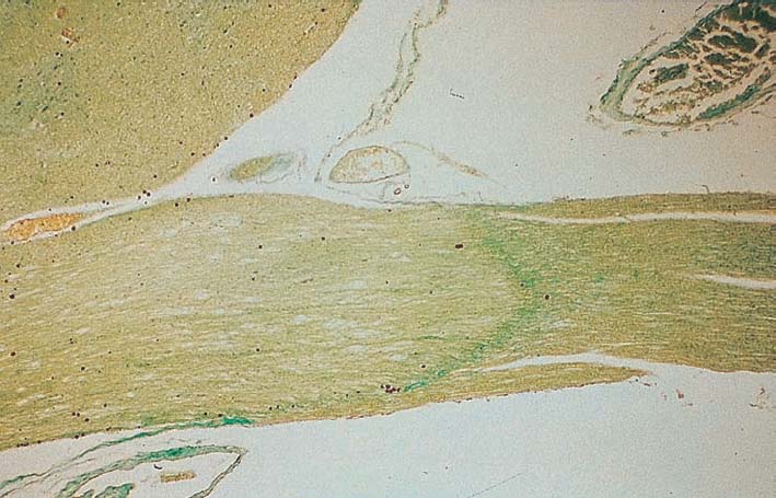
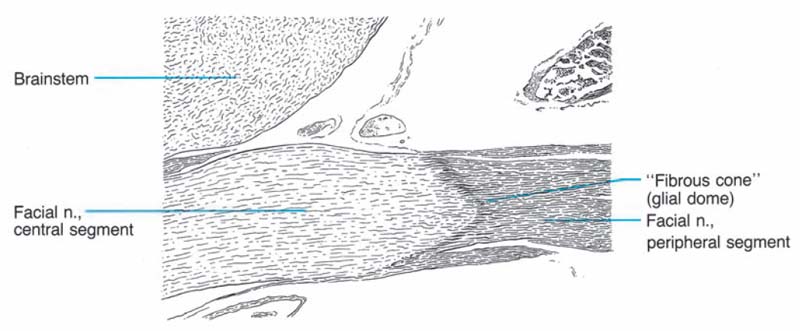
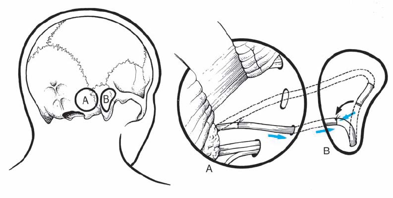
8 Intracranial Reconstruction of the Facial Nerve Fig. 8.1 In this photomicrograph, the border zone between the central myelin and peripheral myelin (Schwann cells) is clearly visualized. In contrast to the eighth nerve, this border is close to the brain stem. This anatomical detail has to be taken into consideration when attempting intracranial reconstruction of the facial nerve. Fig. 8.2 The intracranial–intratemporal facial nerve grafting procedure. The outlined areas are where the bone is removed to expose the seventh nerve at the brain stem and in the mastoid bone. A: right lateral suboccipital craniectomy for exploration and total removal of a cerebellopontine angle tumor. Facial nerve destroyed by the tumor. B: mastoidectomy and opening of the facial nerve canal.
 Neuroanatomy
 Clinical Material
Related posts:


Stay updated, free articles. Join our Telegram channel

Full access? Get Clinical Tree


